Textbook Question
Which of the following is not a modification of a compound light microscope?
a. Brightfield microscopy
b. Darkfield microscopy
c. Electron microscopy
d. Phase-contrast microscopy
e. Fluorescence microscopy
1449
views
 Verified step by step guidance
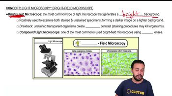
 03:46
03:46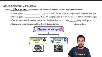 03:08 03:46
Which of the following is not a modification of a compound light microscope?
a. Brightfield microscopy
b. Darkfield microscopy
c. Electron microscopy
d. Phase-contrast microscopy
e. Fluorescence microscopy
Assume that you are viewing a Gram-stained field of red cocci and blue rods through the microscope. You can safely conclude that you have
a. Made a mistake in staining
b. Two different species
c. Old bacterial cells
d. Young bacterial cells
e. None of the above
Assume you stain Clostridium by applying a basic stain, carbolfuchsin, with heat, decolorizing with acid-alcohol, and counterstaining with an acidic stain, nigrosin. Through the microscope, the endospores are 1, and the cells are stained 2.
a. 1—red; 2—black
b. 1—black; 2—colorless
c. 1—colorless; 2—black
d. 1—red; 2—colorless
e. 1—black; 2—red
What is the purpose of a decolorizer in the Gram stain? In the acid-fast stain?
Fill in the following table regarding the Gram stain: