 Back
BackDigestive System: Structure, Function, and Clinical Conditions
Study Guide - Smart Notes
 Tailored notes based on your materials, expanded with key definitions, examples, and context.
Digestive System Overview
The digestive system is responsible for the breakdown, absorption, and metabolism of nutrients, as well as the elimination of waste. It consists of the alimentary canal and accessory organs, each with specialized structures and functions essential for nutrition and health.
Important Vocabulary
Digestion: Breakdown of food materials mechanically or chemically.
Absorption: Passage of nutrients through membranes into the blood.
Metabolism: Complex process by which nutrients are used by the body.
Alimentary canal: Continuous muscular tube of the digestive tract.
Ingestion: Taking food into the GI tract.
Motility: Movement of food through the digestive tract.
Secretion: Release of enzymes and other products into the GI tract.
Elimination: Removal of waste materials from the body.
Feces: Material formed as a result of digestion.
Main and Accessory Organs of the Digestive System
Main organs: Mouth, pharynx, esophagus, stomach, small intestine, large intestine, rectum, anus.
Accessory organs: Teeth, tongue, salivary glands, liver, gallbladder, pancreas.
Primary mechanisms: Ingestion, motility, secretion, digestion (mechanical and chemical), absorption, elimination.
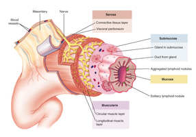
Digestive Tract Wall: Four Layers
The digestive tract wall is composed of four main layers, each with distinct functions that support digestion and absorption.
Mucosa: Innermost layer; mucous membrane open to the outside world, involved in secretion and absorption.
Submucosa: Connective tissue layer containing blood vessels, lymphatics, and nerves; supports the mucosa.
Muscularis: Two to three layers of muscle tissue responsible for motility (movement) of the digestive tract.
Serosa: Outermost covering; a serous membrane (visceral peritoneum) that reduces friction and forms the mesentery.
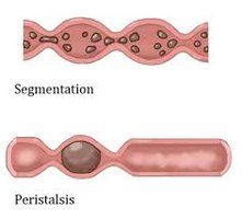
Motility: Segmentation and Peristalsis
Motility refers to the movement of food through the digestive tract, primarily by two mechanisms:
Segmentation: Localized contractions that mix nutrients with digestive juices, enhancing mechanical breakdown.
Peristalsis: Rhythmic, wave-like contractions that propel food along the digestive tract.
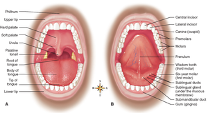
Structures of the Mouth
Oral Cavity
The mouth is the entry point for food and the site where digestion begins. It consists of the oral cavity, teeth, and salivary glands.
Roof: Hard palate (anterior, bone) and soft palate (posterior, muscle); uvula prevents food from entering the nasopharynx.
Floor: Tongue (skeletal muscle, mucous membrane, attached to hyoid bone); frenulum anchors the tongue.
Papillae: Small bumps on the tongue; taste buds are located on the sides of papillae.
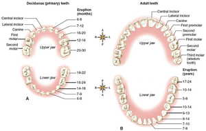
Teeth
Teeth are essential for mechanical digestion, breaking food into smaller pieces to facilitate chemical digestion.
Types: Incisors (cutting), canines (tearing), premolars (crushing), molars (grinding).
Deciduous teeth: 20 baby teeth, usually present by age 2.
Permanent teeth: 32 adult teeth, usually present by age 17-24.
Bolus: Chewed food mixed with saliva, ready for swallowing.
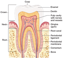
Tooth Structure
Crown: Exposed part, covered by enamel (hardest substance in the body).
Neck: Surrounded by gums (gingiva).
Root: Anchored in jaw bone, held by periodontal membrane.
Salivary Glands
Parotid: Largest, secrete watery enzyme-rich fluid with sodium bicarbonate.
Submandibular: Secrete both enzymes and mucus.
Sublingual: Secrete mostly mucus.
Salivary amylase: Begins chemical digestion of carbohydrates.
Conditions of the Mouth
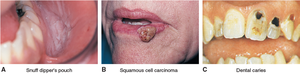
Cancer: Often linked to tobacco and alcohol; leukoplakia and squamous cell carcinoma are common forms.
Dental caries: Tooth decay caused by bacteria and sugar; can lead to cavities and tooth loss.
Gingivitis: Gum inflammation, often due to poor hygiene.
Periodontitis: Advanced gum disease affecting the periodontal membrane and bone.
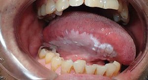
Thrush: Oral candidiasis (yeast infection), common in children and immunocompromised individuals.
Congenital conditions: Cleft lip and palate, due to failure of fusion during development.
Pharynx, Esophagus, and Stomach
Pharynx
Muscular tube behind mouth and nose; part of both digestive and respiratory tracts.
Subdivided into nasopharynx, oropharynx, and laryngopharynx.
Oropharynx is key for swallowing (deglutition).
Esophagus
Muscular, mucus-lined tube (~10 inches) from pharynx to stomach.
Upper esophageal sphincter (UES) prevents air entry; lower esophageal sphincter (LES) prevents reflux.
Conditions of the Esophagus
GERD: Gastroesophageal reflux disease; backward flow of stomach acid.
Hiatal hernia: Stomach protrudes through diaphragm, often causing reflux.
Stomach Structure and Function
Divided into cardia, fundus, body, and pylorus.
Three muscle layers for strong mixing action.
Gastric glands secrete gastric juice (contains HCl and enzymes) and intrinsic factor (for vitamin B12 absorption).
Chyme: Semiliquid food after mixing with gastric juice.
Conditions of the Stomach
Gastritis: Inflammation of the stomach lining.
Ulcers: Open sores, often caused by Helicobacter pylori infection or NSAIDs.
Stomach cancer: Often detected late; risk factors include H. pylori, alcohol, tobacco, and preserved foods.
Small Intestine
Structure
~20 feet long, divided into duodenum, jejunum, and ileum.
Duodenum is the main site for chemical digestion; receives bile and pancreatic juice.
Villi and microvilli increase surface area for absorption.
Function
Primary site for digestion and absorption of nutrients.
Enzymes from intestinal glands and pancreas complete digestion.
Bile emulsifies fats for digestion.
Conditions
Enteritis: Inflammation of the small intestine.
Gastroenteritis: Inflammation of stomach and small intestine.
Malabsorption syndrome: Inadequate absorption of nutrients, leading to various symptoms.
Liver, Gallbladder, and Pancreas
Liver and Gallbladder Structure and Function
Liver: Largest gland, produces bile, metabolizes nutrients, detoxifies substances.
Gallbladder: Stores and concentrates bile, releases it in response to cholecystokinin (CCK).
Bile: Emulsifies fats, aids in cholesterol excretion.
Conditions
Gallstones (cholelithiasis): Solid clumps of cholesterol; may block bile ducts and cause pain or jaundice.
Hepatitis: Inflammation of the liver, caused by viruses, alcohol, or toxins.
Cirrhosis: Chronic liver damage with fibrosis; impairs liver function.
Pancreas
Exocrine function: Secretes digestive enzymes and bicarbonate into the duodenum.
Endocrine function: Secretes insulin and glucagon to regulate blood glucose.
Conditions
Diabetes mellitus: Inadequate insulin production or action.
Pancreatitis: Inflammation due to blocked ducts or enzyme activation within the pancreas.
Pancreatic cancer: Aggressive, poor prognosis.
Cystic fibrosis: Genetic disorder causing thick secretions and blocked pancreatic ducts.
Large Intestine, Appendix, and Peritoneum
Large Intestine Structure and Function
~5 feet long, includes cecum, colon (ascending, transverse, descending, sigmoid), rectum, and anal canal.
Absorbs water and salts, forms feces, houses beneficial bacteria (microbiome).
Bacteria synthesize vitamin K and some B vitamins.
Conditions
Diarrhea: Rapid transit, poor water absorption.
Constipation: Slow transit, excessive water absorption.
Diverticulitis: Inflammation of intestinal pouches (diverticula).
Colitis: Inflammation of the colon; includes Crohn disease and ulcerative colitis.
Colorectal cancer: Malignancy of colon or rectum, often from polyps.
Appendix
Wormlike structure attached to the cecum; incubator for healthy bacteria.
Appendicitis: Inflammation, often requires surgical removal.
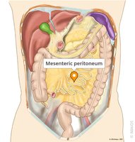
Peritoneum
Serous membrane lining the abdominal cavity and organs.
Parietal layer lines the cavity; visceral layer covers organs.
Peritonitis: Inflammation, often due to infection from ruptured organs.
Ascites: Fluid accumulation in the peritoneal space, often from liver disease.
Mechanical vs. Chemical Digestion
Mechanical digestion: Physical breakdown of food (chewing, mixing, peristalsis).
Chemical digestion: Enzymatic breakdown of macromolecules into absorbable units.
Enzymes: Biological catalysts that facilitate hydrolysis reactions; names often end in -ase.
Digestion of Carbohydrates, Proteins, and Lipids
Carbohydrate Digestion
Begins in the mouth with salivary amylase.
Continues in the small intestine with pancreatic amylase and disaccharidases (maltase, sucrase, lactase).
End products: Monosaccharides (mainly glucose).
Protein Digestion
Begins in the stomach with pepsin (activated by HCl).
Continues in the small intestine with trypsin and peptidases.
End products: Amino acids.
Lipid Digestion
Begins in the small intestine after emulsification by bile.
Pancreatic lipase splits triglycerides into fatty acids and glycerol.
Absorption and Structural Adaptations
Absorption: Movement of nutrients from the intestinal lumen into blood or lymph.
Structural adaptations: Plicae, villi, and microvilli increase surface area for efficient absorption.
Monosaccharides and amino acids are actively transported; fatty acids and glycerol are absorbed into lacteals.
Water follows sodium by osmosis; vitamins are absorbed according to solubility (water- or fat-soluble).
