 Back
BackPeripheral Vascular System: Assessment and Health Promotion
Study Guide - Smart Notes
 Tailored notes based on your materials, expanded with key definitions, examples, and context.
Peripheral Vascular System Overview
Introduction to Peripheral Vascular Health
The peripheral vascular system consists of the arteries, veins, and lymphatic vessels outside the heart and brain. Proper function is essential for tissue perfusion, oxygen delivery, and waste removal. Disorders of this system can lead to significant health problems, including peripheral artery disease (PAD), deep vein thrombosis (DVT), and chronic venous insufficiency.
Key Concepts and Terminology
Essential Terms
Ischemia: Reduced blood flow to tissues, causing oxygen deprivation.
PVD (Peripheral Vascular Disease): Disorders of blood vessels outside the heart and brain.
PAD (Peripheral Artery Disease): A type of PVD involving narrowed arteries, reducing blood flow to limbs.
Embolism: Obstruction of a blood vessel by a clot or foreign material.
DVT (Deep Vein Thrombosis): Formation of a blood clot in a deep vein, usually in the legs.
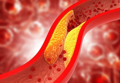
Atherosclerosis: Build-up of fatty deposits (plaques) in arteries, leading to narrowing and reduced blood flow.
Arteriosclerosis: Hardening and loss of elasticity of arterial walls.
Health History and Assessment
Subjective Data Collection
Gathering a thorough health history is crucial for identifying vascular problems. Key questions include:
Presence of leg pain or cramps (claudication distance)
Skin changes on arms or legs
Swelling in legs
Lymph node enlargement
Current medications
Past medical history (e.g., diabetes, hypertension)
Self-care practices
Physical Examination of the Peripheral Vascular System
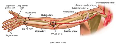
Arteries of the Arm
Major arteries in the arm include the brachial, radial, and ulnar arteries. Pulse sites are important for assessing circulation.
Inspection and Palpation of the Arms
Profile sign: Normal angle < 160°; clubbing suggests chronic hypoxia.
Capillary refill: Normal is less than 2 seconds.
Symmetry: Assess for edema or swelling.
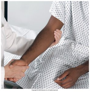
Pulses: Assess radial and brachial pulses for rate, rhythm, elasticity, and amplitude.
Epitrochlear lymph node: Palpate for enlargement.
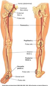
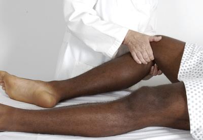
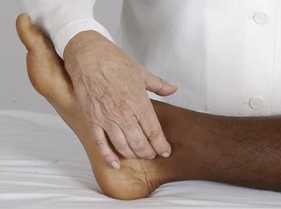
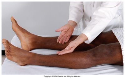
Arteries of the Leg
The main arteries of the leg include the femoral, popliteal, posterior tibial, and dorsalis pedis arteries. Pulse sites are critical for vascular assessment.
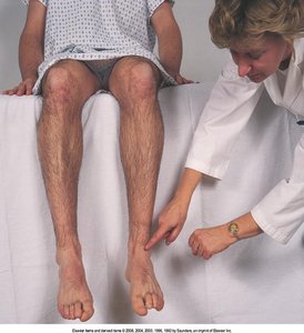
Inspection and Palpation of the Legs
Check for dependent rubor (redness) or elevated pallor (paleness).
Assess hair distribution, especially on toes (loss may indicate poor perfusion).
Observe venous patterns and look for visible, dilated, or tortuous veins.
Measure calf circumference at the widest point for asymmetry.
Inspect for lesions, noting size and location.
Assess temperature (coolness may indicate arterial insufficiency).
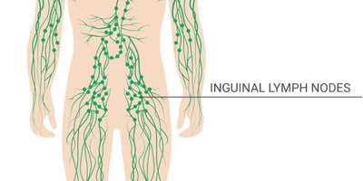
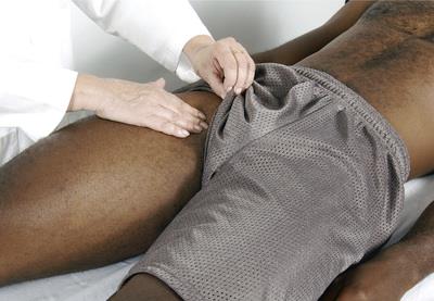
Lymphatic Assessment
Palpate inguinal lymph nodes for enlargement or tenderness.
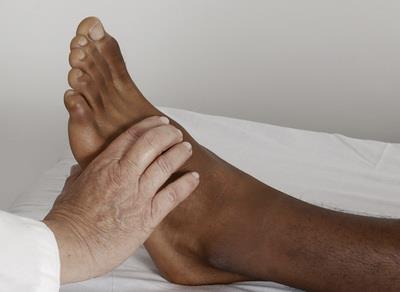
Peripheral Arteries and Pulses in the Leg
Femoral artery: Assess for presence and bruits (abnormal sounds).
Popliteal, posterior tibial, and dorsalis pedis arteries: Palpate for pulses; may be difficult to palpate in older adults.
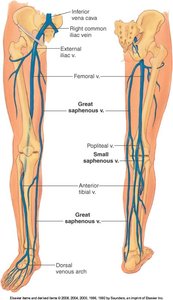
Venous System of the Legs
Major veins include the femoral, popliteal, great and small saphenous veins. Assessment focuses on the presence of varicosities, edema, and venous ulcers.
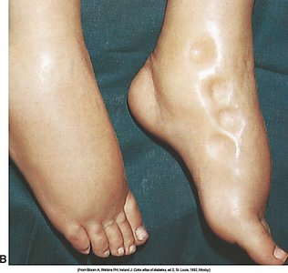
Edema Assessment
Pretibial edema: Depress the tibia or medial malleolus to check for pitting.
Grade pitting edema from 1+ (mild) to 4+ (severe).
Assess for visible, dilated, or tortuous veins.
Evaluate strength and sensation in the lower extremities.
Color Changes and Arterial Deficit
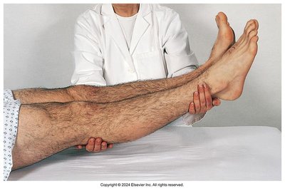
Assess for color return after elevation (normal: < 10 seconds).
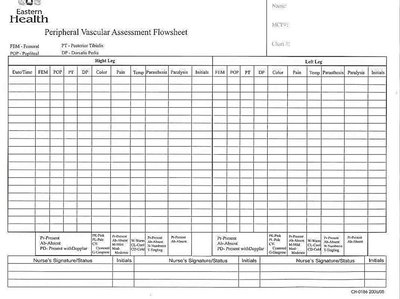
Peripheral Vascular Assessment Checklist
The 6 P's of Acute Limb Ischemia
Pain
Pallor
Paralysis
Paresthesia
Poikilothermia (coolness)
Pulselessness
These signs are critical for identifying acute arterial occlusion.
Diagnostic Tools
Doppler Ultrasonic Stethoscope
A Doppler device magnifies weak pulses, aiding in the detection of blood flow in peripheral arteries when pulses are difficult to palpate.
Developmental Considerations
Infants
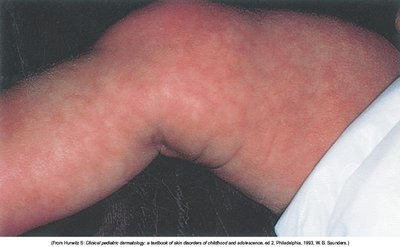
Temporary cyanotic conditions such as transient acrocyanosis and cutis marmorata are common.
Lymph nodes may be palpable.
Pregnant Individuals (Third Trimester)
Pitting edema and varicose veins in the lower legs are common due to increased venous pressure.
Older Adults
Trophic changes (e.g., thin, shiny skin, thick nails, hair loss) may occur.
Posterior tibial and dorsalis pedis pulses may be difficult to palpate.
Health Promotion: Foot Care
Check feet daily for cuts, blisters, or sores.
Promote blood flow by moving and exercising feet regularly.
Wear comfortable, well-fitting shoes.
Keep skin soft and smooth to prevent cracking and infection.
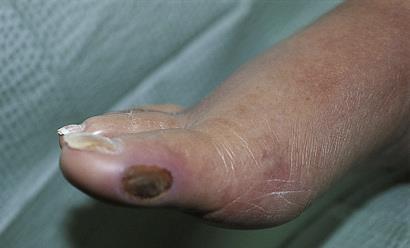
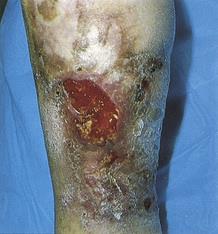
Vascular Insufficiency and Diabetes
Vascular Insufficiency
Chronic arterial or venous insufficiency can lead to ulcers, delayed wound healing, and increased risk of infection, especially in individuals with diabetes.
Case Study Application
Case Study: S.R., 65-year-old Female
Subjective Data to Obtain: Onset, duration, and characteristics of symptoms; history of similar episodes; medication adherence; lifestyle factors.
Pulse Assessment Sites: Radial, brachial, femoral, popliteal, posterior tibial, dorsalis pedis.
Collateral Circulation Evaluation: Allen's test before arterial blood gas sampling.
Likely Cause of Symptoms: Congestive heart failure exacerbation leading to fluid overload, pulmonary congestion, and peripheral edema.
Summary Table: Peripheral Vascular Assessment
Assessment Area | Normal Findings | Abnormal Findings |
|---|---|---|
Capillary Refill | < 2 seconds | > 2 seconds (possible arterial insufficiency) |
Pulses | 2+ (normal amplitude) | 0 (absent), 1+ (weak), 3+ (bounding) |
Edema | None | Pitting (1+ to 4+), non-pitting |
Skin Color | Pink, even | Pallor, cyanosis, rubor |
Hair Distribution | Even on lower extremities | Patchy or absent (arterial disease) |
Additional info: This guide integrates foundational knowledge of peripheral vascular assessment with clinical application, supporting exam preparation and practical skills for personal-health college students.
