 Back
BackSensation and Perception: Visual Stimuli and Color Vision
Study Guide - Smart Notes
 Tailored notes based on your materials, expanded with key definitions, examples, and context.
Visual Stimuli
Nature of Visual Stimuli
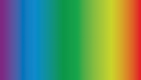
The stimulus for vision is light, specifically electromagnetic energy within the visible spectrum. The properties of light waves determine the qualities we perceive, such as color, brightness, and saturation. The visible light spectrum ranges from approximately 380 to 700 nanometers (nm).
Wavelength: Refers to the distance between successive peaks of a wave. It determines the hue (color) we perceive.
Amplitude: Refers to the height of the wave. It determines the brightness of the light.
Complexity: Refers to the number of different wavelengths present. It determines the saturation (purity or vividness) of the color.
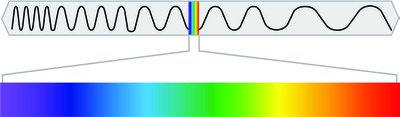
Example: Shorter wavelengths correspond to blue/violet light, while longer wavelengths correspond to red light. Higher amplitude waves appear brighter, and pure wavelengths (single color) are more saturated than mixed wavelengths.
Visual Anatomy
Structure of the Eye
The human eye is a complex organ that focuses light and converts it into neural signals for the brain to interpret.
Cornea: The transparent, protective outer layer that bends (refracts) light as it enters the eye.
Pupil: The opening in the center of the iris that allows light to enter the eye.
Iris: The colored part of the eye that surrounds the pupil and controls its size.
Lens: A transparent structure that changes shape (accommodation) to focus images on the retina.
Retina: The neural tissue lining the back of the eyeball, containing photosensitive cells (rods and cones) that detect light.

Accommodation: The lens changes shape to focus on objects at different distances. With age, the lens loses flexibility, leading to difficulty focusing on close objects (presbyopia).
Pathway of Light: Light passes through the cornea → pupil → lens → retina, where it is converted into neural signals.
Photosensitive Cells
The retina contains two main types of photosensitive cells:
Rods: Rod-shaped cells that respond to low light levels (scotopic vision). They are located mainly in the periphery of the retina and are numerous. Rods are responsible for detecting movement and are not sensitive to color.
Cones: Cone-shaped cells that respond to bright light (photopic vision). They are concentrated in the fovea (center of the retina) and are fewer in number. Cones are responsible for color vision and high visual acuity. There are three types of cones, each sensitive to different wavelengths (short, medium, long).

Fovea: The central region of the retina with a high density of cones, providing sharp central vision.
Blind Spot: The point where the optic nerve exits the eye; there are no rods or cones here, resulting in a small gap in the visual field.
Mnemonic: Cones see in Color, near the Center, and are sCarce.
Color Vision
Trichromatic Theory
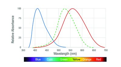
The trichromatic theory explains the first stage of color vision, which occurs in the retina. According to this theory, there are three types of cones, each sensitive to a different range of wavelengths (short/blue, medium/green, long/red). The combination of activation across these cones allows us to perceive a wide range of colors.
Short-wavelength cones: Most sensitive to blue light (~420 nm).
Medium-wavelength cones: Most sensitive to green light (~534 nm).
Long-wavelength cones: Most sensitive to red light (~564 nm).
Limitation: The trichromatic theory cannot explain all aspects of color vision, such as afterimages and certain types of colorblindness.
Opponent Process Theory
The opponent process theory describes the second stage of color processing, which occurs in the nervous system (beyond the retina). This theory proposes that color perception is controlled by the activity of two opponent systems: red-green, blue-yellow, and black-white. Opponent process cells are excited by one color and inhibited by its opponent.
Red/Green
Blue/Yellow
Black/White
Afterimage: A phenomenon where staring at a color for a prolonged period causes you to see its opponent color when you look away. For example, staring at a green image may produce a red afterimage.
This pattern of activation is interpreted by the visual cortex, allowing for the perception of a full range of colors.
Colorblindness
Colorblindness occurs when one or more types of cones are nonfunctional. The most common type is deuteranopia (green cone deficiency). Individuals with deuteranopia have difficulty distinguishing between red and green hues.
Well-seen colors: Blue and red (depending on which cones are functional).
Poorly seen colors: Green (if green cones are nonfunctional).
Application: Colorblindness tests and adaptive technologies help individuals with color vision deficiencies navigate the world more effectively.
Comparison of Color Vision Theories
Theory | Main Idea | Explains | Limitation |
|---|---|---|---|
Trichromatic Theory | Three types of cones sensitive to different wavelengths | Basic color mixing, color matching | Cannot explain afterimages |
Opponent Process Theory | Opponent pairs of colors processed by opponent cells | Afterimages, color contrast effects | Does not explain initial color detection |
Summary Table: Properties of Light and Perception
Wave Property | Perceived Property |
|---|---|
Wavelength | Hue (Color) |
Amplitude | Brightness |
Complexity | Saturation |
Additional info: These notes cover the foundational aspects of visual sensation and perception, focusing on the physical properties of light, the anatomy of the eye, the function of rods and cones, and the major theories of color vision. Understanding these concepts is essential for further study in biological psychology and perception.
