 Back
BackSensation and Perception: Visual Stimuli and Color Vision
Study Guide - Smart Notes
 Tailored notes based on your materials, expanded with key definitions, examples, and context.
Ch. 4 Sensation and Perception
Visual Stimuli
The process of vision begins with the detection of visual stimuli, specifically light waves, by the eye. The properties of these light waves determine the qualities of the visual experience.
Stimulus for Vision: The primary stimulus for vision is light, specifically electromagnetic energy within the visible spectrum (approximately 380 to 700 nm).
Wave Properties and Perceived Qualities:
Wavelength: The distance between successive peaks of a wave; determines hue (color).
Amplitude: The height of the wave; determines brightness.
Complexity: The number of different wavelengths present; determines saturation (purity of color).
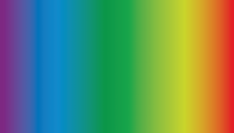
Visible Light Spectrum: The portion of the electromagnetic spectrum that is visible to the human eye, ranging from violet (short wavelength) to red (long wavelength).
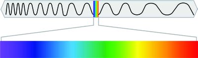
Example: The visible spectrum can be illustrated as a range of colors from violet to red, each corresponding to a different wavelength.
Practice: Matching Wave and Perceived Properties
Wavelength is associated with hue.
Amplitude is associated with brightness.
Complexity is associated with saturation.
Visual Anatomy
Structure of the Eye
The human eye is a complex organ that focuses light and converts it into neural signals for the brain to interpret.
Cornea: The transparent, protective outer layer that bends light as it enters the eye.
Pupil: The opening in the center of the iris that allows light to enter the eye.
Iris: The colored part of the eye that surrounds the pupil and controls its size.
Lens: A transparent structure that changes shape (accommodation) to focus light on the retina.
Retina: The neural tissue lining the back of the eyeball; contains photosensitive cells (rods and cones) responsible for vision.
Accommodation: The process by which the lens changes shape to focus on objects at different distances.

Example: Light enters the eye through the cornea, passes through the pupil (controlled by the iris), is focused by the lens, and finally reaches the retina where it is converted into neural signals.
Practice: Age and Accommodation
As people age, the lens loses flexibility, reducing the ability to accommodate and often requiring reading glasses.
Photosensitive Cells in the Retina
The retina contains two main types of photosensitive cells: rods and cones, each with distinct functions.
Rods:
Rod-shaped cells that respond to dim light.
Responsible for achromatic vision (black and white).
Located primarily in the periphery of the retina.
Numerous in number.
Detect movement and are sensitive to low light levels.
Cones:
Cone-shaped cells that respond to bright light.
Responsible for color vision.
Concentrated in the fovea (center of the retina).
Fewer in number compared to rods.
Three types, each sensitive to different wavelengths (red, green, blue).
Fovea: Area of the retina with the highest concentration of cones; responsible for sharp central vision.
Blind Spot: The point where the optic nerve exits the eye; contains no rods or cones.

Example: Looking at a colorful painting relies on cones, while seeing in dim light or detecting movement in the periphery relies on rods.
Color Vision
Trichromatic Theory
The trichromatic theory explains color vision at the level of the retina, where three types of cones respond to different wavelengths of light.
Three Types of Cones: Each type is sensitive to either short (blue), medium (green), or long (red) wavelengths.
First Stage of Color Processing: Occurs in the retina.
Limitation: Trichromatic theory cannot explain all aspects of color vision, such as afterimages.
Example: Mixing red, green, and blue light in different proportions can produce any color perceived by humans.
Opponent Process Theory
The opponent process theory describes a second stage of color processing that occurs in the nervous system, beyond the retina.
Opponent Pairs: Colors are processed in opposing pairs: red/green, blue/yellow, and black/white.
Opponent Process Cells: These cells are excited by one color and inhibited by its opponent.
Afterimages: Prolonged viewing of one color can lead to the perception of its opponent color when looking away.
Example: Staring at a red image for a long time and then looking at a white surface may produce a green afterimage.
Colorblindness
Colorblindness is a condition in which one or more types of cones are nonfunctional, affecting color perception.
Deuteranopia: The most common type, where the green-sensitive cones are nonfunctional.
Effects: Individuals with deuteranopia have difficulty distinguishing between red and green hues.
Example: A person with deuteranopia can see blues and reds well but struggles with greens.
Summary Table: Properties of Light and Perception
Wave Property | Perceived Property |
|---|---|
Wavelength | Hue (Color) |
Amplitude | Brightness |
Complexity | Saturation |
Summary Table: Rods vs. Cones
Property | Rods | Cones |
|---|---|---|
Shape | Rod-shaped | Cone-shaped |
Light Sensitivity | Dim light | Bright light |
Color Vision | No | Yes |
Location | Periphery | Fovea (center) |
Number | Many | Few |
Additional info: The notes above expand on the original content by providing definitions, examples, and summary tables for clarity and completeness.
