 Back
BackAnatomy & Physiology Exam 3 Study Guide – Muscles and Nervous System
Study Guide - Smart Notes
 Tailored notes based on your materials, expanded with key definitions, examples, and context.
Q1. How are muscles classified based on their actions and shapes? What are the key anatomical features of skeletal muscle?
Background
Topic: Muscle Classification and Skeletal Muscle Anatomy
This question tests your understanding of how muscles are grouped by their function (actions) and physical appearance (shapes), as well as your knowledge of the basic structure of skeletal muscle tissue.
Key Terms:
Muscle actions: The specific movements a muscle produces (e.g., flexion, extension, abduction).
Muscle shapes: Descriptions such as fusiform, pennate, circular, convergent, and parallel.
Skeletal muscle anatomy: Includes muscle fibers, fascicles, connective tissue layers (epimysium, perimysium, endomysium), and the neuromuscular junction.
Step-by-Step Guidance
List the main ways muscles are classified by their actions (e.g., agonist, antagonist, synergist, fixator).
Describe the different muscle shapes and give an example of each (e.g., deltoid is triangular, biceps brachii is fusiform).
Identify the major anatomical structures of a skeletal muscle, including the connective tissue coverings and arrangement of muscle fibers.
Relate how the structure of a muscle can influence its function and movement.
Try solving on your own before revealing the answer!
Q2. Where are the following muscles located in the body, and what are their primary functions? (Deltoid, biceps, pectoralis, serratus, sternocleidomastoid, rotator cuff muscles, triceps, hamstrings group, masseter, abdominal muscles, quadriceps group, gastrocnemius, soleus)
Background
Topic: Major Muscles of the Human Body
This question assesses your ability to identify the location and function of key muscles, which is essential for understanding movement and muscle actions.
Key Terms:
Origin and insertion: Where a muscle begins and ends on the skeleton.
Muscle function: The movement produced when the muscle contracts.
Step-by-Step Guidance
For each muscle listed, identify its anatomical location (e.g., deltoid is on the shoulder).
Describe the primary action or function of each muscle (e.g., biceps brachii flexes the elbow).
Group muscles by region (e.g., upper limb, lower limb, head/neck, trunk) to help organize your study.
Try solving on your own before revealing the answer!
Q3. What is a muscle lever system, and how does it relate to movement?
Background
Topic: Biomechanics of Muscle Action
This question focuses on the concept of levers in the body, which helps explain how muscles and bones interact to produce movement.
Key Terms and Formulas:
Lever: A rigid bar (bone) that moves on a fixed point (fulcrum) when a force (muscle contraction) is applied.
Types of levers: First-class, second-class, and third-class levers.
Mechanical advantage: $\text{Mechanical Advantage} = \frac{\text{Effort Arm}}{\text{Load Arm}}$
Step-by-Step Guidance
Define the components of a lever system: fulcrum, effort, and load.
Describe the three classes of levers and give an example of each in the human body.
Explain how the arrangement of these components affects the force and speed of movement.
Try solving on your own before revealing the answer!
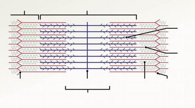
Q4. What is the structure of a sarcomere, and what are its key microscopic features?
Background
Topic: Muscle Cell (Fiber) Structure
This question tests your knowledge of the microscopic anatomy of muscle, specifically the sarcomere, which is the functional unit of muscle contraction.
Key Terms:
Sarcomere: The repeating unit of a myofibril, defined by Z-discs.
Key features: Z-disc, M-line, A-band, I-band, H-zone, actin (thin filament), myosin (thick filament).
Step-by-Step Guidance
Draw or label a diagram of a sarcomere, identifying the Z-discs, A-band, I-band, H-zone, and M-line.
Describe the arrangement of actin and myosin filaments within the sarcomere.
Explain how the structure of the sarcomere relates to its function in muscle contraction.
Try solving on your own before revealing the answer!
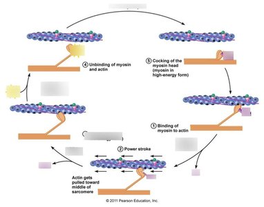
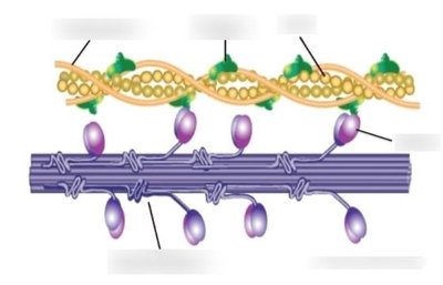
Q5. What is the sliding filament theory of muscle contraction?
Background
Topic: Physiology of Muscle Contraction
This question examines your understanding of how muscles contract at the molecular level, specifically how actin and myosin interact.
Key Terms:
Sliding filament theory: The process by which myosin heads pull actin filaments toward the center of the sarcomere, shortening the muscle.
Cross-bridge cycle: The repeated formation and breaking of bonds between actin and myosin.
Step-by-Step Guidance
Describe the role of myosin and actin filaments in muscle contraction.
Explain the sequence of events in the cross-bridge cycle (attachment, power stroke, detachment, re-cocking).
Discuss the role of ATP in the contraction and relaxation process.
Try solving on your own before revealing the answer!
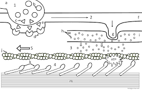
Q6. What are the main steps in the sequence of muscle contraction events?
Background
Topic: Excitation-Contraction Coupling
This question focuses on the sequence of events from the nerve signal to muscle contraction, including the role of the neuromuscular junction.
Key Terms:
Excitation-contraction coupling: The process linking the action potential in a muscle fiber to contraction.
Neuromuscular junction: The synapse between a motor neuron and a muscle fiber.
Step-by-Step Guidance
Describe the arrival of the action potential at the neuromuscular junction and the release of neurotransmitter (acetylcholine).
Explain how the action potential spreads along the sarcolemma and into the T-tubules.
Discuss the release of calcium ions from the sarcoplasmic reticulum and their role in initiating contraction.
Try solving on your own before revealing the answer!
Q7. What is the difference between isometric and isotonic muscle contractions?
Background
Topic: Types of Muscle Contraction
This question tests your understanding of the two main types of muscle contractions and their physiological significance.
Key Terms:
Isometric contraction: Muscle tension increases, but the muscle does not change length.
Isotonic contraction: Muscle changes length (shortens or lengthens) while tension remains constant.
Step-by-Step Guidance
Define isometric and isotonic contractions and provide an example of each.
Explain the physiological conditions under which each type occurs.
Discuss the functional significance of each type in daily activities.
Try solving on your own before revealing the answer!
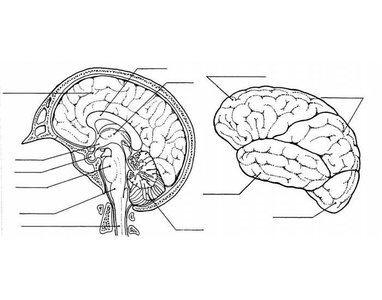
Q8. What are the general structures and functions of the nervous system? What are the main types of nervous cells?
Background
Topic: Nervous System Anatomy and Physiology
This question covers the organization of the nervous system and the different types of cells involved in its function.
Key Terms:
Central nervous system (CNS): Brain and spinal cord.
Peripheral nervous system (PNS): Nerves outside the CNS.
Neurons: Nerve cells that transmit signals.
Neuroglia: Supporting cells in the nervous system.
Step-by-Step Guidance
Describe the main divisions of the nervous system (CNS and PNS) and their primary functions.
List the major types of cells in the nervous system and their roles (neurons, astrocytes, oligodendrocytes, Schwann cells, etc.).
Explain how the structure of neurons supports their function in signal transmission.
Try solving on your own before revealing the answer!
