 Back
BackAnatomy & Physiology Exam 3 Study Guide – Muscles and Nervous System
Study Guide - Smart Notes
 Tailored notes based on your materials, expanded with key definitions, examples, and context.
Q1. How are muscles classified based on their actions and shapes? What are the key anatomical features of skeletal muscle?
Background
Topic: Muscle Classification and Skeletal Muscle Anatomy
This question tests your understanding of how muscles are grouped by their function (actions) and physical appearance (shapes), as well as your knowledge of the basic structure of skeletal muscle tissue.
Key Terms:
Muscle actions: The specific movements a muscle produces (e.g., flexion, extension, abduction).
Muscle shapes: Descriptions such as fusiform (globe), parallel, convergent(deltoid), pennate, circular, etc.
Skeletal muscle anatomy: Includes muscle fibers, fascicles, connective tissue layers (epimysium, perimysium, endomysium), and the neuromuscular junction.
Step-by-Step Guidance
List the main ways muscles are classified by action (e.g., agonist, antagonist, synergist, fixator) and by shape (e.g., deltoid = triangular, biceps = fusiform).
Describe the general structure of a skeletal muscle, including the arrangement of muscle fibers into fascicles and the connective tissue coverings.
Identify the major anatomical features visible in a typical diagram of skeletal muscle (such as the sarcolemma, myofibrils, and nuclei).
Try solving on your own before revealing the answer!
Q2. Where are the following muscles located in the body, and what are their primary functions? (Deltoid, biceps, pectoralis, serratus, sternocleidomastoid, rotator cuff muscles, triceps, hamstrings group, masseter, abdominal muscles, quadriceps group, gastrocnemius, soleus)
Background
Topic: Major Muscles of the Human Body
This question assesses your ability to identify the location and function of key muscles, which is essential for understanding movement and muscle anatomy.
Key Terms:
Origin and insertion: Where a muscle starts and attaches on the skeleton.
Muscle function: The movement produced when the muscle contracts.
Step-by-Step Guidance
For each muscle listed, identify its general location (e.g., deltoid = shoulder, biceps = upper arm).
Briefly describe the main action or function of each muscle (e.g., biceps brachii = flexes the elbow).
Group muscles by region (e.g., upper limb, lower limb, head/neck, trunk) to help organize your study.
Try solving on your own before revealing the answer!
Q3. What is a muscle lever system, and how does it relate to movement?
Background
Topic: Biomechanics of Muscle Action
This question explores how bones and muscles interact as levers to produce movement, a key concept in understanding body mechanics.
Key Terms and Formulas:
Lever: A rigid bar (bone) that moves on a fixed point (fulcrum) when a force (muscle contraction) is applied.
Types of levers: First-class, second-class, third-class (most common in the body).
Mechanical advantage:
Step-by-Step Guidance
Define the components of a lever system: fulcrum, effort, and load.
Describe the three classes of levers and give an example of each from the human body.
Explain how the arrangement of these components affects the force and speed of movement.
Try solving on your own before revealing the answer!
Q4. What is the microscopic structure of a sarcomere, and how does it relate to muscle contraction?
Background
Topic: Sarcomere Anatomy and Function
This question focuses on the organization of the sarcomere, the basic contractile unit of muscle, and its role in muscle contraction.
Key Terms:
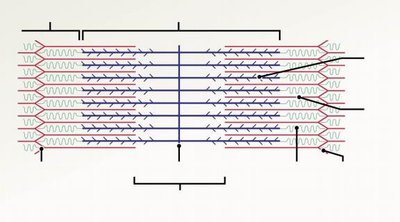
Sarcomere: The segment between two Z lines in a myofibril.
Myofilaments: Actin (thin) and myosin (thick) filaments.
Bands and lines: A band, I band, H zone, M line, Z disc.
Step-by-Step Guidance
Label the main structures of a sarcomere on a diagram (Z disc, M line, A band, I band, H zone).
Describe the arrangement of thick and thin filaments within the sarcomere.
Explain how the sliding of these filaments leads to muscle contraction.
Try solving on your own before revealing the answer!
Q5. Explain the sliding filament theory of muscle contraction.
Background
Topic: Muscle Physiology – Sliding Filament Theory
This question tests your understanding of how actin and myosin filaments interact to produce muscle contraction.
Key Terms:
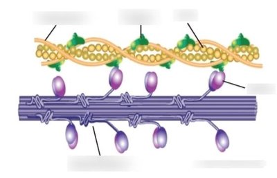
Actin: Thin filament protein.
Myosin: Thick filament protein with heads that bind to actin.
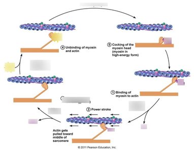
Cross-bridge cycle: The repeated formation and breaking of bonds between actin and myosin.
Step-by-Step Guidance
Describe the role of calcium ions and ATP in initiating contraction.
Explain how myosin heads bind to actin to form cross-bridges.
Outline the sequence of events in the cross-bridge cycle (attach, pivot, detach, reset).
Try solving on your own before revealing the answer!
Q6. What is the sequence of events in muscle contraction?
Background
Topic: Muscle Contraction Sequence
This question asks you to describe the step-by-step process from nerve impulse to muscle fiber contraction.
Key Terms:
Neuromuscular junction: The synapse between a motor neuron and a muscle fiber.
Action potential: Electrical signal that triggers contraction.
Excitation-contraction coupling: The link between the action potential and muscle contraction.
Step-by-Step Guidance
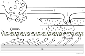
Describe the arrival of the nerve impulse at the neuromuscular junction and the release of neurotransmitter (acetylcholine).
Explain how the action potential spreads along the sarcolemma and into the T-tubules.
Describe the release of calcium from the sarcoplasmic reticulum and its effect on the actin-myosin interaction.
Try solving on your own before revealing the answer!
Q7. What is the difference between isometric and isotonic muscle contractions?
Background
Topic: Types of Muscle Contraction
This question tests your understanding of how muscles generate force with or without changing length.
Key Terms:
Isometric contraction: Muscle generates force without changing length.
Isotonic contraction: Muscle changes length while generating force (can be concentric or eccentric).
Step-by-Step Guidance
Define isometric and isotonic contractions with examples.
Explain the physiological conditions under which each type occurs.
Describe how muscle tension and length change in each type.
Try solving on your own before revealing the answer!
Q8. Describe the general structure and functions of the nervous system, including synapses, neuron polarization, depolarization, repolarization, and hyperpolarization.
Background
Topic: Nervous System Structure and Function
This question covers the organization of the nervous system and the basic physiology of nerve signaling.
Key Terms:
Neuron: Nerve cell that transmits electrical signals.
Synapse: Junction between two neurons or a neuron and a muscle cell.
Polarization: Resting state of the neuron membrane.
Depolarization: Membrane potential becomes less negative.
Repolarization: Return to resting membrane potential.
Hyperpolarization: Membrane potential becomes more negative than resting.
Step-by-Step Guidance
Describe the main divisions of the nervous system (central and peripheral) and their functions.
Explain the structure of a typical neuron and the role of synapses in communication.
Outline the sequence of events during an action potential, including polarization, depolarization, repolarization, and hyperpolarization.
Try solving on your own before revealing the answer!
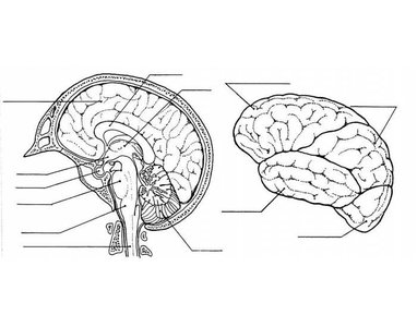
Q9. Identify and describe the functions of the following nervous system structures: cerebral cortex, spinocerebellar tracts, arbor vitae, brain stem, hypothalamus, primary auditory cortex, meninges, ventricles, CSF, gray and white matter, somatosensory and somatomotor cortex, motor neurons, limbic system, diencephalon, basal nuclei.
Background
Topic: Central Nervous System Anatomy and Function
This question requires you to locate and describe the function of major brain and nervous system structures.
Key Terms:
Cerebral cortex: Outer layer of the brain, involved in higher functions.
Spinocerebellar tracts: Pathways that carry proprioceptive information to the cerebellum.
Arbor vitae: White matter of the cerebellum.
Brain stem: Controls basic life functions.
Hypothalamus: Regulates homeostasis.
Primary auditory cortex: Processes sound information.
Meninges: Protective coverings of the brain and spinal cord.
Ventricles: Cavities in the brain that contain cerebrospinal fluid (CSF).
Gray matter: Contains neuron cell bodies.
White matter: Contains myelinated axons.
Somatosensory cortex: Processes sensory input.
Somatomotor cortex: Controls voluntary movement.
Motor neurons: Carry signals to muscles.
Limbic system: Involved in emotion and memory.
Diencephalon: Includes thalamus and hypothalamus.
Basal nuclei: Involved in movement regulation.
Step-by-Step Guidance
Locate each structure on a brain diagram and briefly describe its function.
Group structures by region (e.g., cerebrum, cerebellum, brainstem, diencephalon) for easier memorization.
Relate each structure to its role in sensory processing, motor control, or homeostasis.
Try solving on your own before revealing the answer!
