 Back
BackBlood Vessels and Circulation: Structure, Function, and Regulation
Study Guide - Smart Notes
 Tailored notes based on your materials, expanded with key definitions, examples, and context.
Blood Vessels and Circulation
Introduction to Blood Vessels and Circulation
The cardiovascular system relies on a network of blood vessels to transport blood throughout the body. Blood vessels are classified by size and histological organization, and they play a critical role in regulating blood flow and pressure. The largest vessels, the pulmonary trunk and aorta, are directly attached to the heart and initiate the pulmonary and systemic circuits, respectively.
Types and Structure of Blood Vessels
Classification of Blood Vessels
Arteries: Carry blood away from the heart.
Arterioles: Small branches of arteries leading to capillary beds.
Capillaries: Smallest vessels; site of exchange between blood and interstitial fluid.
Venules: Smallest branches of veins collecting blood from capillaries.
Veins: Return blood to the heart.
Layers of Vessel Walls
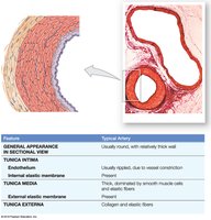
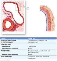
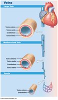
Tunica intima: Inner layer; includes endothelium and connective tissue. In arteries, contains an internal elastic membrane.
Tunica media: Middle layer; composed of smooth muscle and elastic fibers. In arteries, separated from the externa by an external elastic membrane.
Tunica externa: Outer layer; contains collagen and elastic fibers, and in veins, smooth muscle cells. Large vessels contain vasa vasorum (small vessels that supply the vessel wall).
Differences Between Arteries and Veins
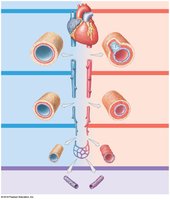
Arteries have thicker walls and higher blood pressure than veins.
Arteries have a small, round lumen; veins have a large, irregular lumen.
Arteries are more elastic; veins have valves to prevent backflow.
Types of Arteries
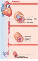
Elastic arteries: Large vessels (e.g., aorta); tunica media rich in elastic fibers to absorb pressure waves.
Muscular arteries: Medium-sized; tunica media contains many muscle cells for distribution.
Arterioles: Smallest arteries; little or no tunica externa, thin or incomplete tunica media.
Types of Veins
Venules: Collect blood from capillaries.
Medium-sized veins: Thin tunica media, few muscle cells, tunica externa with elastic fibers.
Large veins: All three tunics present; thick externa, thin media.
Venous valves: Folds of tunica intima that prevent backflow; failure leads to varicose veins or hemorrhoids.
Capillaries: Structure and Function
Capillary Types
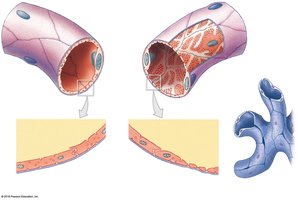
Continuous capillaries: Complete endothelial lining; found in most tissues; restricts passage of cells and proteins.
Fenestrated capillaries: Have pores; allow rapid exchange of water and solutes; found in kidneys, endocrine glands, intestines.
Sinusoids: Large gaps between endothelial cells; allow passage of large proteins and cells; found in liver, spleen, bone marrow.

Capillary Beds and Blood Flow Regulation
Capillary beds connect arterioles and venules.
Precapillary sphincters regulate blood flow into capillaries.
Thoroughfare channels provide direct connections between arterioles and venules.
Anastomoses are connections between vessels that provide alternate routes for blood flow.
Angiogenesis is the formation of new blood vessels, important in development and response to hypoxia.
Distribution and Dynamics of Blood Flow
Blood Distribution
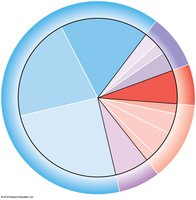
Heart, arteries, and capillaries contain 30–35% of blood volume.
Venous system contains 65–70% of blood volume; acts as a blood reservoir.
Pressure, Resistance, and Blood Flow
Blood flow (F) is determined by the pressure gradient (ΔP) and resistance (R):
Blood pressure (BP): Arterial pressure, measured in mm Hg.
Capillary hydrostatic pressure (CHP): Pressure within capillary beds.
Venous pressure: Pressure in the venous system.
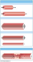
Total peripheral resistance is affected by vessel length, diameter, blood viscosity, and turbulence.
Cardiovascular Pressures and Blood Velocity
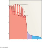
As blood moves from arteries to capillaries, pressure and velocity decrease, while total cross-sectional area increases.
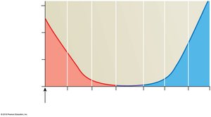
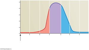
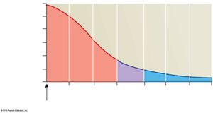
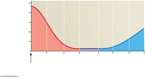
Blood Pressure Terms
Systolic pressure: Peak arterial pressure during ventricular contraction.
Diastolic pressure: Minimum arterial pressure during relaxation.
Pulse pressure: Difference between systolic and diastolic pressure.
Mean arterial pressure (MAP):
Capillary Exchange: Mechanisms and Forces
Mechanisms of Exchange
Diffusion: Movement of substances from high to low concentration.
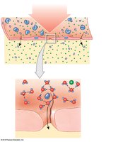
Filtration: Driven by hydrostatic pressure; water and small solutes move out of capillaries.
Reabsorption: Driven by osmotic pressure; water moves into capillaries due to higher solute concentration in blood.
Net Filtration Pressure (NFP)
Determines direction of fluid movement:
At arterial end: Filtration dominates (fluid out).
At venous end: Reabsorption dominates (fluid in).
Excess fluid enters lymphatic vessels.
Regulation of Blood Flow and Pressure
Local, Neural, and Endocrine Control
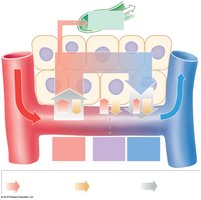
Autoregulation: Immediate, local adjustments via precapillary sphincters.
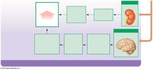
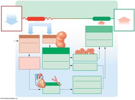
Neural mechanisms: Cardiovascular centers in the medulla oblongata adjust cardiac output and vessel diameter.
Endocrine mechanisms: Hormones (e.g., E, NE, ADH, angiotensin II, EPO, ANP, BNP) regulate blood volume and pressure.
Baroreceptor and Chemoreceptor Reflexes
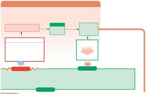
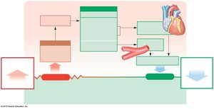
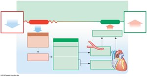
Baroreceptors: Detect changes in blood pressure; located in carotid sinuses, aortic sinuses, and right atrium.
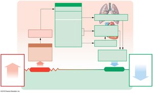
Chemoreceptors: Monitor pH, O2, and CO2 levels; located in carotid and aortic bodies, and medulla oblongata.
Special Circulations and Adaptations
Special Circulations
Brain: Maintains constant blood flow; cerebral vessels dilate if peripheral vessels constrict.
Heart: Coronary arteries supply blood; increased demand leads to vasodilation.
Lungs: Blood flow regulated by O2 levels in alveoli.
Exercise and Hemorrhage
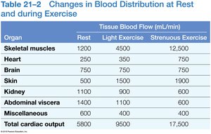
Light exercise increases vasodilation, venous return, and cardiac output.
Heavy exercise maximizes cardiac output, restricts blood flow to nonessential organs, and redirects it to muscles, lungs, and heart.
Hemorrhage triggers short-term (neural, hormonal) and long-term (fluid recall, erythropoiesis) responses to restore blood pressure and volume.
Pulmonary and Systemic Circuits
Pulmonary Circuit
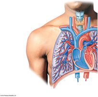
Deoxygenated blood travels from right ventricle to lungs via pulmonary arteries.
Gas exchange occurs in pulmonary capillaries; oxygenated blood returns to left atrium via pulmonary veins.
Systemic Circuit
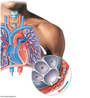
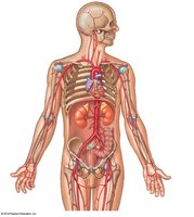
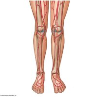
Supplies oxygenated blood to all body tissues except the lungs.
Major arteries branch from the aorta; major veins return blood to the right atrium via the superior and inferior vena cava.
Fetal and Maternal Circulation
Fetal Circulation Adaptations
Placenta provides O2 and nutrients; umbilical arteries and vein transport blood between fetus and placenta.
Foramen ovale and ductus arteriosus allow blood to bypass fetal lungs.
At birth, these shunts close, and normal pulmonary circulation begins.
Congenital Heart Defects
Patent foramen ovale: Foramen ovale remains open, causing left-to-right shunt.
Patent ductus arteriosus: Ductus arteriosus remains open, causing right-to-left shunt and cyanosis.
Tetralogy of Fallot: Combination of four defects including pulmonary stenosis and ventricular septal defect.
Ventricular septal defect: Opening in interventricular septum; most common congenital heart defect.
Atrioventricular septal defect: Incomplete separation of atria and ventricles; often associated with Down syndrome.
Transposition of the great vessels: Aorta and pulmonary artery are switched.
Effects of Aging and System Integration
Age-Related Changes in the Cardiovascular System
Decreased hematocrit, increased risk of thrombus, pooling of blood in legs due to valve deterioration.
Reduced cardiac output, changes in conduction system, increased atherosclerosis, and scar tissue formation in the heart.
Arteries become less elastic, increased risk of aneurysm, calcium and lipid deposits, and increased risk of stroke or infarction.
Integration with Other Body Systems
The cardiovascular system interacts with all other organ systems, transporting nutrients, hormones, and wastes, regulating pH and temperature, and defending against pathogens.
