 Back
BackBones and Bone Structure: Study Notes for ANP College Students (Ch. 6)
Study Guide - Smart Notes
 Tailored notes based on your materials, expanded with key definitions, examples, and context.
Bones and Bone Structure
An Introduction to Bone and Skeletal Tissue
The skeletal system is composed of bones and associated organs such as cartilage. Bone is a living, dynamic tissue that responds to its environment and plays several essential roles in the body.
Support: Bones provide structural support for the body.
Protection: Bones protect soft internal organs (e.g., the skull protects the brain, ribs protect the heart and lungs).
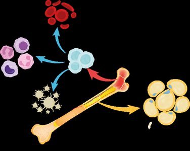
Production of Blood Cells: Bones produce blood cells through hematopoiesis in the bone marrow.
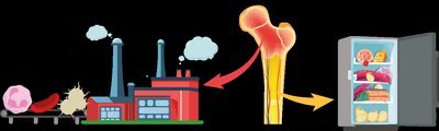
Storage: Bones store fat (triglycerides) and minerals, especially calcium.
Levers for Muscles: Bones act as levers for muscle action, enabling movement.


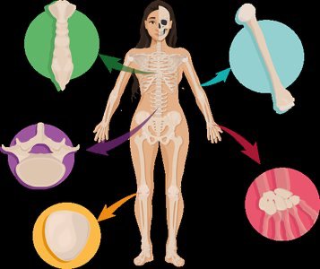
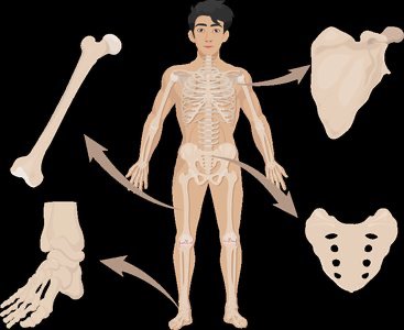
Types of Bones
Bones are categorized based on their shape, which relates to their function and location in the body.
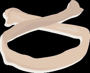
Long Bones: Shaped like a shaft with expanded ends. Examples: Arm and leg bones.
Short Bones: Cube-shaped. Examples: Wrist and ankle bones.
Flat Bones: Thin, flat, and slightly curved. Examples: Sternum, ribs, cranial bones.
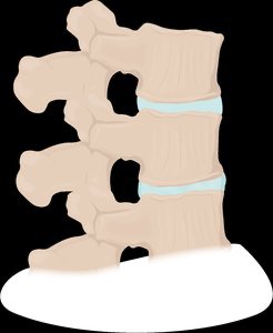
Irregular Bones: Complex shapes. Examples: Pelvis, facial bones.
Sesamoid Bones: Develop inside a tendon, variable in number. Example: Patella.

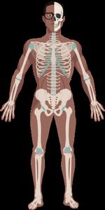
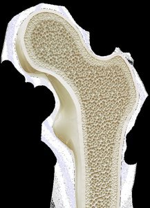
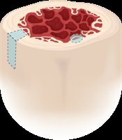
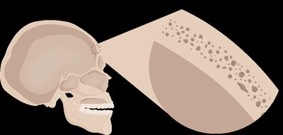
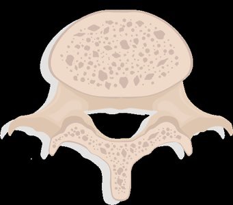
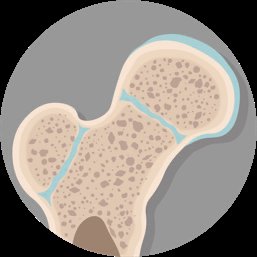
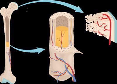
Gross Anatomy of Bone: Compact and Spongy Bone
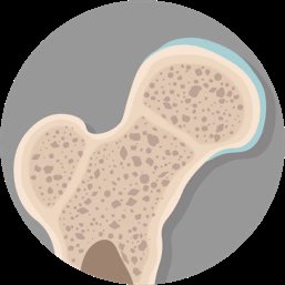
Bones have two main structural arrangements: compact bone and spongy bone. These arrangements provide strength and flexibility.
Compact Bone: Appears solid, with no visible spaces. Optimized for strength and hardness. Found at the edges of all bones and in the shaft of long bones.
Spongy Bone: Looks like a sponge, built like scaffolding. Contains trabeculae (bone struts) and reduces weight. Spaces are filled with marrow and found in the middle of bones.

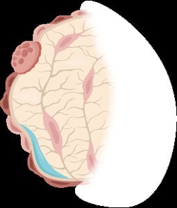
Gross Anatomy of Bone: Periosteum and Endosteum
Connective tissue layers surround and line bones, providing blood, nerves, attachment points, and bone stem cells.
Periosteum: Covers the outside surface of bone; consists of two layers (fibrous and osteogenic). Vascular and innervated. Collagen from tendons and ligaments weave into the fibrous layer. Perforating fibers (collagen) enter the bone matrix.
Endosteum: Lines all inner surfaces of bone and has the same composition as the osteogenic layer.


Gross Anatomy of Bone: Bone Marrow
Bone marrow fills the spaces inside bones and is crucial for blood cell production and fat storage.
Red Marrow: Site of hematopoiesis (formation of blood cells). Found in spongy bone in adults and is the primary marrow type in babies.
Yellow Marrow: Stores triglycerides (fat). Found in spongy bone and medullary cavity. Primary marrow type in adults but can revert to red marrow if needed.

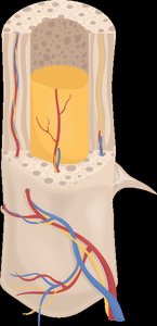
Gross Anatomy of Bone: Structure of a Long Bone
Long bones consist of two ends (epiphyses) and a shaft (diaphysis), with a medullary cavity inside the shaft.
Epiphysis: Wider end of a long bone, covered with articular cartilage at the joint.
Diaphysis: Tubular shaft of the bone, contains yellow marrow in the medullary cavity.
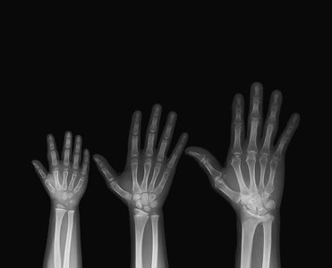
Metaphysis: Area where epiphysis and diaphysis meet; contains the epiphyseal plate (site of bone growth until puberty).
Epiphyseal Line: Plate converts to compact bone when growth is done.

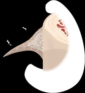
Gross Anatomy of Bone: Nerves and Blood Supply
Bones contain blood vessels and nerves, which enter through the nutrient foramen.
Nutrient Foramen: Small hole in the diaphysis for blood vessels and nerves.
Nutrient Artery: Brings blood into the medullary cavity.
Nutrient Vein: Carries blood out of the bone.
Nerves: Pass through the nutrient foramen into the bone.
Microscopic Anatomy of Bones: Bone Matrix
The extracellular matrix of bone has two basic components: inorganic and organic.
Inorganic Matrix: Hydroxyapatite crystals (calcium and phosphate), making up about 2/3 of bone mass. Provides hardness.
Organic Matrix (Osteoid): Collagen fibers and ground substance, making up about 1/3 of bone mass. Provides strength and flexibility.
Formula: Hydroxyapatite:

Microscopic Anatomy of Bones: Bone Cells
Bone tissue (osseous tissue) is comprised of cells and extracellular matrix. The main bone cells are:
Osteoprogenitor (Osteogenic) Cells: Stem cells of the periosteum and endosteum that give rise to osteoblasts.
Osteoblasts: Build bone by secreting bone matrix (collagen fibers and calcium-binding enzymes). Mature osteoblasts become osteocytes.
Osteocytes: Mature bone cells that maintain the matrix. Housed in lacunae, monitor bone stress, and contribute to calcium homeostasis. Cellular projections allow communication via gap junctions.
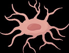
Osteoclasts: Break down bone matrix for remodeling. Multinucleate, derived from white blood cells, have a ruffled border to increase surface area, secrete acid and enzymes to dissolve inorganic and organic matrix.

Microscopic Anatomy of Bones: The Osteon
The osteon (Haversian system) is the structural unit of compact bone, comprised of concentric rings (lamellae) of matrix.
Central Canal: Contains blood vessels and nerves, runs parallel to the length of the bone.
Perforating Canals: Run perpendicular to central canals, connect central canal to other blood vessels and nerves.
Canaliculi: Small channels in all directions, allow communication and transport between osteocytes.
Lacunae: Chambers that contain osteocytes.
Microscopic Anatomy of Bones: Structure of Lamellae
Collagen fibers within lamellae run in alternate directions, increasing bone strength and resistance to twisting forces.
Within Lamella: Collagen runs in the same direction, providing strength.
Between Adjacent Lamellae: Collagen runs in alternate directions, resisting twisting.
Microscopic Anatomy of Bones: Trabeculae
Spongy bone is named for its sponge-like appearance with many open spaces. Trabeculae are small rods or struts in spongy bone, containing lamellae, osteocytes, and canaliculi. They align with lines of stress and do not have well-organized osteons or central canals.
Trabecular Bone: Also called cancellous bone; trabeculae means beam or timber.
Canaliculi: Allow osteocytes to receive nutrients in spongy bone.
Summary Tables
Summary Table: Bone Types and Features
Bone Type | Shape | Example | Main Function |
|---|---|---|---|
Long | Shaft, expanded ends | Femur, humerus | Leverage, movement |
Short | Cube-shaped | Carpals, tarsals | Stability, support |
Flat | Thin, flat, curved | Sternum, ribs, skull | Protection |
Irregular | Complex shape | Vertebrae, pelvis | Protection, support |
Sesamoid | Within tendon | Patella | Reduce friction |
Summary Table: Bone Cells and Functions
Cell Type | Function | Location |
|---|---|---|
Osteoprogenitor | Stem cell, forms osteoblasts | Periosteum, endosteum |
Osteoblast | Builds bone matrix | Bone surface |
Osteocyte | Maintains matrix | Lacunae |
Osteoclast | Breaks down matrix | Bone surface |
Additional info: Academic context and explanations were expanded for clarity and completeness. All images included are directly relevant to the adjacent content and reinforce key concepts.
