 Back
BackBones and Bone Tissue: Structure, Function, and Physiology
Study Guide - Smart Notes
 Tailored notes based on your materials, expanded with key definitions, examples, and context.
Bones and Bone Tissue
The Skeletal System: Overview
The skeletal system is a complex organ system that includes bones, joints, and supporting tissues. Bones are the main organs, with adults typically having 206 bones. Each bone is composed of osseous tissue, dense regular and irregular connective tissue, and bone marrow.
Bones: Provide structure and support.
Joints: Allow movement and flexibility.
Bone Marrow: Site of blood cell formation and fat storage.
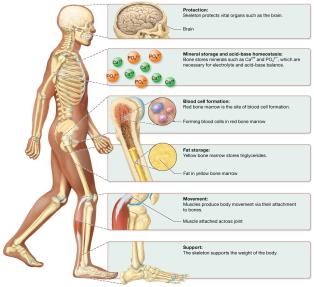
Functions of the Skeletal System
Bones serve several essential functions for the human body:
Protection: Bones such as the skull, sternum, and ribs protect vital organs.
Mineral Storage & Acid-Base Homeostasis: Bones store minerals (calcium, phosphorus, magnesium) critical for electrolyte and acid-base balance.
Blood Cell Formation: Red bone marrow is the site of hematopoiesis (formation of blood cells).
Fat Storage: Yellow bone marrow stores triglycerides in adipocytes.
Movement: Bones act as levers for muscle action, enabling movement.
Support: The skeleton provides structural framework and supports body weight.
Bone Structure
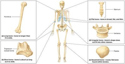
Classification of Bones by Shape
Bones are classified based on their shape, which relates to their function and location in the body:
Long Bones: Longer than wide (e.g., humerus, femur).
Short Bones: About as long as wide, cube-shaped (e.g., wrist, ankle bones).
Flat Bones: Thin and broad (e.g., skull, pelvis).
Irregular Bones: Irregular shapes (e.g., vertebrae).
Sesamoid Bones: Small, flat, oval-shaped, within tendons (e.g., patella).
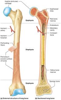
Structure of a Long Bone
Long bones have a distinct anatomy that supports their function:
Periosteum: Dense irregular connective tissue membrane covering the bone, containing blood vessels and nerves.
Perforating Fibers: Collagen anchors that attach periosteum to bone matrix.
Diaphysis: Shaft of the bone, containing the medullary (marrow) cavity lined by endosteum.
Epiphyses: Ends of the bone, filled with red marrow and covered with articular cartilage (hyaline cartilage).
Compact Bone: Dense outer layer resisting compression and twisting.
Spongy Bone: Inner honeycomb-like structure, housing bone marrow.
Epiphyseal Lines: Remnants of growth plates (hyaline cartilage) in adults.
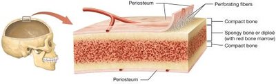
Structure of Short, Flat, Irregular, and Sesamoid Bones
These bones share similarities with long bones but have fewer structures. In flat bones, the spongy bone is called diploë, and some skull bones contain sinuses to reduce weight.
Bone Marrow
Bone marrow exists in two forms:
Red Bone Marrow: Site of hematopoiesis; abundant in children, limited to certain bones in adults.
Yellow Bone Marrow: Contains adipocytes and blood vessels; increases with age.
The Extracellular Matrix of Bone
Inorganic and Organic Matrix
The bone matrix is composed of inorganic and organic components:
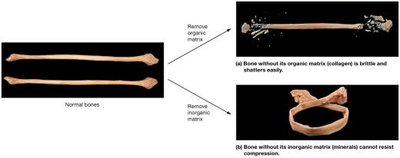
Inorganic Matrix: ~65% of bone weight; mainly hydroxyapatite crystals (calcium and phosphate), providing strength and resistance to compression.
Organic Matrix (Osteoid): ~35% of bone weight; includes collagen fibers, proteoglycans, glycosaminoglycans, glycoproteins, and osteocalcin, providing flexibility and resistance to tension.
Bone Cells
Types of Bone Cells
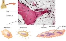
Bone is a dynamic tissue, constantly remodeled by three main cell types:
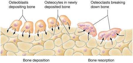
Osteoblasts: Build bone by secreting matrix; derived from osteogenic cells.
Osteocytes: Mature osteoblasts trapped in lacunae; maintain bone matrix.
Osteoclasts: Large, multinucleated cells that break down bone matrix (bone resorption).
Functions of Osteoblasts and Osteocytes
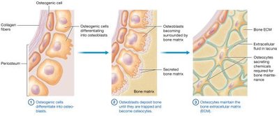
Osteoblasts deposit bone matrix and become osteocytes, which maintain the matrix and recruit osteoblasts for repair.
Function of Osteoclasts
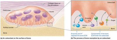
Osteoclasts break down bone ECM by secreting hydrogen ions and enzymes, releasing minerals and organic components into the blood.
Histology of Bone
Compact Bone
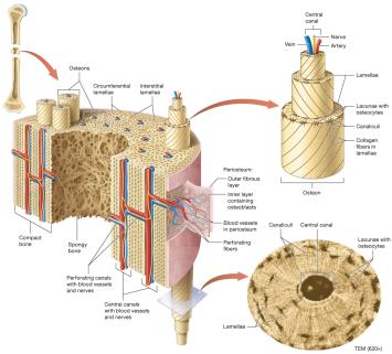
Compact bone is organized into osteons (Haversian systems):
Lamellae: Concentric rings of bone matrix.
Central (Haversian) Canal: Contains blood vessels and nerves.
Lacunae: Small cavities housing osteocytes.
Canaliculi: Tiny canals connecting lacunae for nutrient exchange.
Interstitial and Circumferential Lamellae: Strengthen bone.
Perforating (Volkmann) Canals: Connect osteons and carry blood vessels.
Spongy Bone
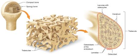
Spongy bone consists of trabeculae, which house osteocytes and allow access to blood supply from bone marrow.
Bone Formation: Ossification
Ossification (Osteogenesis)
Ossification is the process of bone formation, occurring in two main forms:
Intramembranous Ossification: Forms flat bones from a mesenchymal membrane.
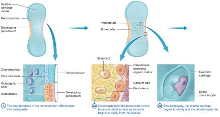
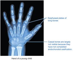
Endochondral Ossification: Forms long and short bones from a hyaline cartilage model.
Steps of Intramembranous Ossification
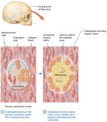
Osteoblasts develop in the primary ossification center from mesenchymal cells.
Osteoblasts secrete organic matrix, which calcifies; trapped osteoblasts become osteocytes.
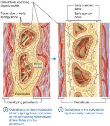
Osteoblasts lay down trabeculae of early spongy bone; some mesenchyme becomes periosteum.
Osteoblasts in periosteum lay down early compact bone; fontanels in newborns are areas of incomplete fusion.
Steps of Endochondral Ossification
Chondroblasts in perichondrium differentiate into osteoblasts.
Osteoblasts build bone collar on external surface; internal cartilage calcifies and chondrocytes die.
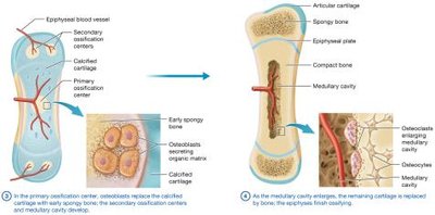
Osteoblasts replace calcified cartilage with early spongy bone; secondary ossification centers and medullary cavity develop.
Remaining cartilage is replaced by bone; epiphyses finish ossifying; cartilage remains in epiphyseal plates and articular cartilage.
Bone Growth
Longitudinal Growth
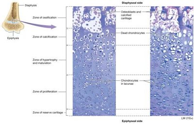
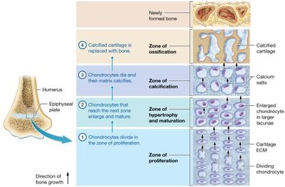
Long bones grow in length at the epiphyseal plate, which has five zones:
Zone of Reserve Cartilage: Cells not directly involved in growth.
Zone of Proliferation: Actively dividing chondrocytes.
Zone of Hypertrophy and Maturation: Mature chondrocytes.
Zone of Calcification: Dead, calcified chondrocytes.
Zone of Ossification: Osteoblasts build bone.
Appositional Growth
Appositional growth increases bone width by osteoblasts laying down new bone between periosteum and bone surface.
The Role of Hormones in Bone Growth
Growth Hormone: Increases mitosis of chondrocytes and osteogenic cells, stimulates osteoblasts.
Testosterone: Increases appositional growth and mitosis, accelerates epiphyseal plate closure.
Estrogen: Similar effects, but less pronounced; epiphyseal plates close earlier in females.
Bone Remodeling
Bone Deposition and Resorption
Bone remodeling is a continual process involving deposition by osteoblasts and resorption by osteoclasts. It maintains calcium homeostasis, repairs bone, and adapts to stress.
Factors Influencing Bone Remodeling
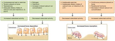
Hormones: Testosterone promotes deposition; estrogen inhibits osteoclasts.
Age: Hormone levels decline, reducing bone formation.
Nutrient Intake: Calcium, vitamin D, K, C, and protein are essential for bone health.
Calcium Ion Homeostasis
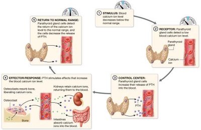
Calcium ions are vital for muscle contraction, nerve transmission, and blood clotting. Blood calcium is regulated by negative feedback loops:
Parathyroid Hormone (PTH): Increases blood calcium by stimulating bone resorption.
Calcitonin: Decreases blood calcium by promoting bone deposition.
Bone Repair
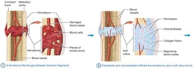
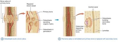
Steps of Fracture Healing
Hematoma Formation: Blood vessels rupture, forming a hematoma.
Soft Callus Formation: Fibroblasts and chondroblasts produce connective tissue and cartilage.
Bone Callus Formation: Osteoblasts lay down primary bone.
Bone Remodeling: Primary bone is replaced with secondary bone.
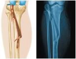
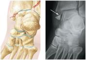
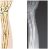
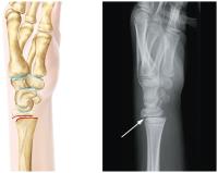
Types of Fractures
Fractures are classified based on their characteristics:
Fracture Type | Description |
|---|---|
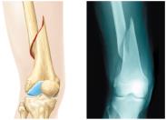
Spiral | Resulting from twisting forces; diaphysis is dislocated. |
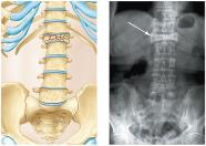
Compression | Bone is crushed under weight; common in vertebrae. |
Comminuted | Bone is shattered into multiple fragments. |
Avulsion | Tendon or ligament pulls off a fragment of bone. |
Greenstick | Bone breaks on one side, bends on the other; common in children. |
Epiphyseal Plate | Involves the growth plate; may interfere with growth. |
Summary Table: Bone Cell Types
Cell Type | Function |
|---|---|
Osteoblast | Builds bone matrix |
Osteocyte | Maintains bone matrix |
Osteoclast | Breaks down bone matrix |
Key Equations
Hydroxyapatite Formula:
Additional info: These notes expand on the original slides and images, providing definitions, examples, and academic context for each topic. All images included are directly relevant to the adjacent content and reinforce key concepts in bone structure, function, and pathology.
