 Back
BackCell Structure, Function, and the Cell Cycle: ANP College Study Guide
Study Guide - Smart Notes
 Tailored notes based on your materials, expanded with key definitions, examples, and context.
Cellular Structure and Function
Cytoplasm and Organelles
The cytoplasm is the region between the plasma membrane and the nucleus, containing cytosol, inclusions, and organelles. Organelles are specialized structures that perform distinct cellular functions, and are classified as either membranous or nonmembranous.
Cytosol: Gel-like fluid with dissolved proteins, salts, and sugars.
Inclusions: Insoluble molecules such as glycogen granules, pigments, lipid droplets, and crystals.
Organelles: Metabolic machinery of the cell, including mitochondria, endoplasmic reticulum, Golgi apparatus, lysosomes, peroxisomes, ribosomes, cytoskeleton, and centrioles.
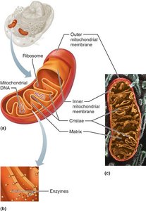
Mitochondria
Mitochondria are the "powerhouses" of the cell, generating ATP through aerobic respiration. They are enclosed by a double membrane, with the inner membrane forming cristae that house proteins essential for cellular respiration. Mitochondria contain their own DNA, RNA, and ribosomes, and divide by fission.
Structure: Outer and inner membranes, cristae, matrix.
Function: ATP production via aerobic respiration.
Unique Features: Own genetic material and bacterial-like division.
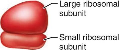
Ribosomes
Ribosomes are nonmembranous organelles responsible for protein synthesis. They consist of protein and ribosomal RNA (rRNA) and exist in two forms: free ribosomes (synthesize proteins for cytosol or organelles) and membrane-bound ribosomes (attached to rough ER, synthesize proteins for membranes, lysosomes, or export).
Structure: Large and small subunits.
Function: Protein synthesis and folding.
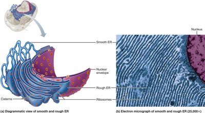
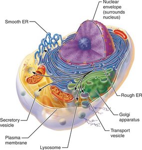
Endoplasmic Reticulum (ER)
The ER is a network of membranous tubules continuous with the nuclear envelope. It is divided into rough ER (RER) and smooth ER (SER).
Rough ER: Studded with ribosomes; synthesizes proteins for secretion, membrane, and lysosomes.
Smooth ER: Lacks ribosomes; involved in lipid metabolism, steroid synthesis, detoxification, glycogen breakdown, and calcium storage.
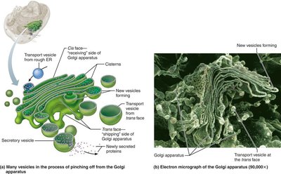
Golgi Apparatus
The Golgi apparatus consists of stacked, flattened membranous sacs (cisterns). It modifies, concentrates, and packages proteins and lipids received from the rough ER. Proteins are sorted into vesicles for secretion, membrane incorporation, or lysosomal digestion.
Steps: Transport vesicles fuse to cis face, proteins are modified, sorted, and packaged at trans face.
Vesicle Types: Secretory vesicles, membrane vesicles, lysosomal vesicles.
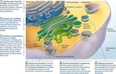
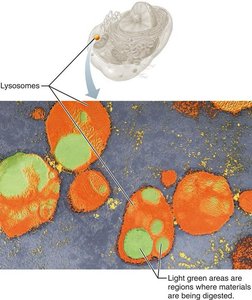
Lysosomes
Lysosomes are spherical membranous sacs containing digestive enzymes (acid hydrolases). They digest bacteria, viruses, toxins, and nonfunctional organelles, and play a role in autolysis and metabolic functions such as glycogen breakdown.
Function: Intracellular digestion and recycling.
Clinical Example: Tay-Sachs disease results from defective lysosomal enzymes, leading to neurodegeneration.
Peroxisomes
Peroxisomes are membranous sacs containing enzymes that detoxify harmful substances and neutralize free radicals. They use oxidase and catalase to convert toxins to hydrogen peroxide and then to water, and are involved in fatty acid metabolism.
Function: Detoxification and lipid metabolism.
The Endomembrane System
The endomembrane system includes the ER, Golgi apparatus, secretory vesicles, lysosomes, nuclear envelope, and plasma membrane. These structures work together to produce, degrade, store, and export biological molecules, and to degrade harmful substances.
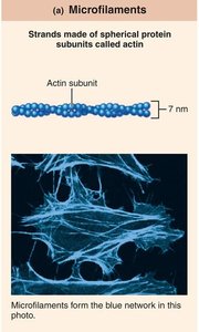
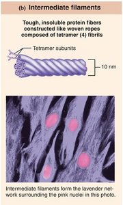
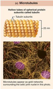
Cytoskeleton
The cytoskeleton is an elaborate network of protein rods that provide structural support, shape, and movement for the cell. It consists of microfilaments, intermediate filaments, and microtubules.
Microfilaments: Thinnest, made of actin; involved in cell shape, motility, and membrane support.
Intermediate Filaments: Medium thickness, ropelike; provide tensile strength and resist pulling forces.
Microtubules: Largest, hollow tubes of tubulin; determine cell shape, organelle distribution, and serve as tracks for motor proteins.
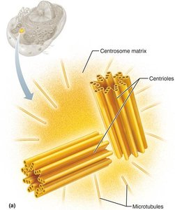
Centrosome and Centrioles
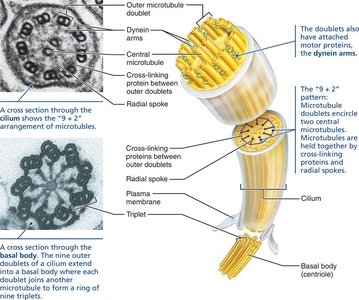
The centrosome is the microtubule organizing center near the nucleus, containing a pair of barrel-shaped centrioles. Centrioles generate microtubules, organize the mitotic spindle, and form the bases of cilia and flagella.
Structure: Pinwheel array of nine triplets of microtubules.
Function: Cell division and formation of cellular extensions.
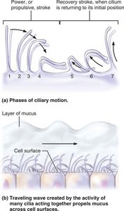
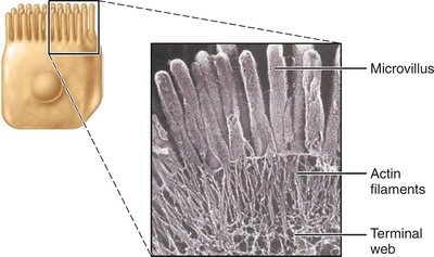
Cilia, Flagella, and Microvilli
Cilia and flagella are cellular extensions that aid in movement. Cilia move substances across cell surfaces, while flagella propel cells (e.g., sperm). Microvilli increase surface area for absorption and contain actin filaments for structural support.
Cilia: "9 + 2" microtubule pattern, coordinated movement.
Flagella: Longer, single extension for cell propulsion.
Microvilli: Fingerlike projections with actin core.
Nucleus and Genetic Material
Nucleus
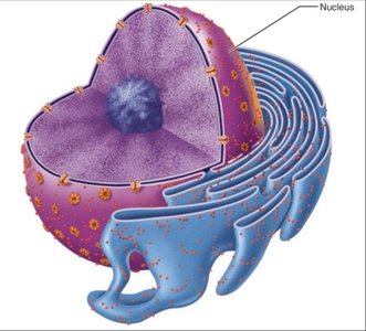
The nucleus is the largest organelle, containing the genetic library for protein synthesis. It responds to cellular signals and regulates gene expression. Most cells are uninucleate, but some are multinucleate or anucleate.
Structures: Nuclear envelope, nucleoli, chromatin.
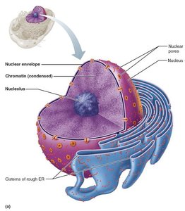
Nuclear Envelope
The nuclear envelope is a double-membrane barrier enclosing the nucleoplasm. The outer layer is continuous with rough ER and studded with ribosomes, while the inner layer (nuclear lamina) maintains nuclear shape. Nuclear pores regulate transport of molecules.
Nucleoli
Nucleoli are spherical bodies within the nucleus involved in rRNA synthesis and ribosome assembly. Usually, one or two are present per nucleus.
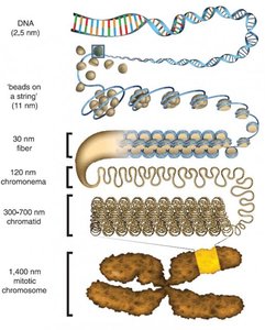
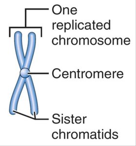
Chromatin and Chromosomes
Chromatin is composed of DNA, histone proteins, and RNA. It is organized into nucleosomes (DNA wrapped around histones). Chromosomes are condensed chromatin, protecting genetic material during cell division.
Composition: 30% DNA, 60% histone proteins, 10% RNA.
Function: Regulation of gene expression and protection during division.
The Cell Cycle and DNA Replication
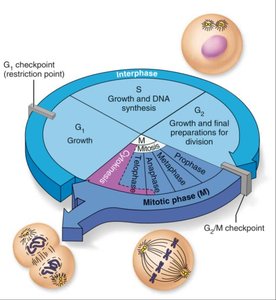
Cell Cycle
The cell cycle is the series of events from cell formation to reproduction. It consists of interphase (growth and preparation) and the mitotic phase (cell division).
Interphase: G1 (growth), S (DNA synthesis), G2 (preparation for division).
Mitotic Phase: Mitosis (nuclear division) and cytokinesis (cytoplasmic division).
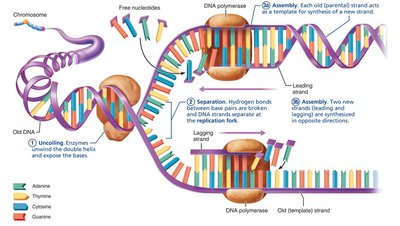
DNA Replication
DNA replication occurs during the S phase of interphase, ensuring each daughter cell receives a complete set of genetic information. The process is semiconservative, with each new DNA molecule containing one old and one new strand.
Steps: Uncoiling, separation, assembly (leading and lagging strands), and splicing.
Enzymes: DNA polymerase (synthesis), DNA ligase (splicing).
Mitosis
Mitosis is the process of nuclear division, consisting of prophase, metaphase, anaphase, and telophase. Cytokinesis divides the cytoplasm, resulting in two daughter cells.
Prophase: Chromatin condenses, spindle forms, nuclear envelope breaks down.
Metaphase: Chromosomes align at the metaphase plate.
Anaphase: Sister chromatids separate and move to opposite poles.
Telophase: Chromosomes uncoil, nuclear membranes reform.
Cytokinesis: Actin ring forms cleavage furrow, cells split.
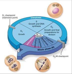
Control of Cell Division
Cell division is regulated by internal and external factors, including cell size, chemical signals, and contact inhibition. Checkpoints ensure proper division and repair of errors.
G1 Checkpoint: Restriction point; if not passed, cell enters G0 (no division).
Genetic Code and Protein Synthesis
Genes and Genetic Code
A gene is a segment of DNA coding for a polypeptide. The genetic code is determined by the sequence of nitrogen bases, organized in triplets. Exons code for amino acids, while introns are noncoding regions.
Triplet Code: Three sequential bases specify an amino acid.
Codons: mRNA sequences corresponding to DNA triplets.
Anticodons: tRNA sequences complementary to mRNA codons.
RNA Types and Roles
Messenger RNA (mRNA): Carries genetic code from DNA to ribosomes.
Ribosomal RNA (rRNA): Structural component of ribosomes.
Transfer RNA (tRNA): Brings amino acids to ribosomes, matches codons with anticodons.
Protein Synthesis: Transcription and Translation
Protein synthesis occurs in two steps: transcription (DNA to mRNA) and translation (mRNA to polypeptide).
Transcription: DNA code is copied to mRNA in the nucleus.
Translation: mRNA is decoded at the ribosome, tRNA brings amino acids, and polypeptide is assembled.
Genetic Code Table
Codon | Amino Acid |
|---|---|
Ala | Alanine |
Arg | Arginine |
Asn | Asparagine |
Asp | Aspartic acid |
Cys | Cysteine |
Glu | Glutamic acid |
Gln | Glutamine |
Gly | Glycine |
His | Histidine |
Ile | Isoleucine |
Leu | Leucine |
Lys | Lysine |
Met | Methionine |
Phe | Phenylalanine |
Pro | Proline |
Ser | Serine |
Thr | Threonine |
Trp | Tryptophan |
Tyr | Tyrosine |
Val | Valine |
Apoptosis
Apoptosis is programmed cell death, essential for removing unneeded, damaged, or infected cells. It involves activation of caspases, degradation of DNA and cytoskeleton, and phagocytosis of cell remnants.
Importance: Maintains tissue health and prevents cancer.
Cell Differentiation and Growth
All cells contain the same DNA, but differentiation occurs through selective gene expression. Cell division is required for growth, repair, and maintenance. Hyperplasia increases cell numbers, while atrophy decreases cell size due to loss of stimulation or use.
Key Equations and Concepts
DNA Replication:
Central Dogma:
Genetic Code:
Additional info: This guide expands on brief points with academic context, definitions, and examples to ensure completeness and clarity for ANP college students.
