 Back
BackCellular Structure, Function, and Division: Study Notes for Anatomy & Physiology
Study Guide - Smart Notes
 Tailored notes based on your materials, expanded with key definitions, examples, and context.
Cell Theory and Contributors
Historical Foundations of Cell Biology
The cell theory is a fundamental concept in biology, stating that all living things are composed of cells, the cell is the basic unit of structure and function, and all cells arise from preexisting cells. Key contributors include Robert Hooke (first described cells), Van Leeuwenhoek (studied living cells), Schleiden and Schwann (formulated cell theory), and Singer & Nicolson (fluid mosaic model of cell membrane).
Cell Theory: All living things are made of cells; cells are the basic unit of life; cells come from preexisting cells.
Microscopic Anatomy: Cytology (study of cells) and Histology (study of tissues).
Types of Cells: Prokaryotic vs. Eukaryotic
Comparative Structure and Function
Cells are classified as prokaryotic (lacking a true nucleus, e.g., bacteria) or eukaryotic (possessing a nucleus, e.g., animal and plant cells). Eukaryotic cells are larger and more complex, with multiple linear chromosomes and membrane-bound organelles.
Feature | Prokaryotic | Eukaryotic |
|---|---|---|
Size | Small | Large |
Complexity | No | Yes |
Ribosomes | Yes (smaller) | Yes (larger) |
Nuclear Membrane | No (nucleoid) | Yes |
Chromosomes | One, coiled | Many, linear |
Cell Wall | Yes (bacteria) | Yes (plants) |
DNA | One, coiled | Many, linear |
Animal vs. Plant Cells
Feature | Animal Cell | Plant Cell |
|---|---|---|
Vacuole | Small | Large |
Produces Food | No | Yes |
Chloroplast | No | Yes |
Cell Wall | No | Yes |
Cell Diversity, Shapes, and Sizes
Variety of Cell Types and Functions
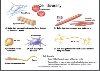
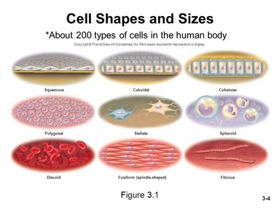
Human body contains over 200 types of cells, differing in shape, size, and function. These differences are essential for specialized roles in tissues and organs.
Columnar: Long and narrow (e.g., lining of intestines)
Cuboidal: Equal dimensions (e.g., kidney tubules)
Discoid: Disc-shaped (e.g., erythrocytes)
Stellate: Star-shaped (e.g., neurons)
Fusiform: Spindle-shaped (e.g., muscle cells)
Spherical: Round (e.g., leukocytes)
Structure of a Typical Animal Cell
Main Components and Their Functions
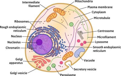
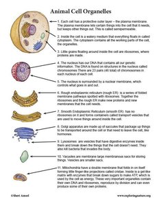
A typical animal cell consists of three main parts: nucleus, cytoplasm, and cell membrane. Each part contains specialized structures (organelles) that perform distinct functions.
Nucleus: Largest organelle, contains genetic material (DNA), surrounded by nuclear envelope.
Cytoplasm: Gel-like substance containing organelles; site of metabolic activity.
Cell Membrane: Selectively permeable barrier, regulates entry and exit of substances.
Nucleus: Structure and Function
Genetic Control Center
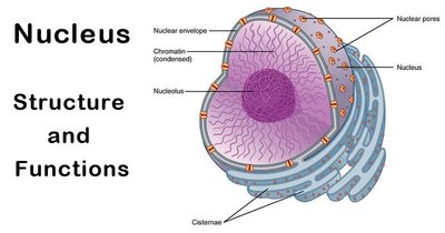
The nucleus is the control center of the cell, housing DNA and coordinating activities such as growth, metabolism, and reproduction. It is surrounded by a double membrane with nuclear pores for transport.
Nuclear Envelope: Double membrane with pores for exchange between nucleus and cytoplasm.
Nucleolus: Site of ribosome synthesis, contains RNA.
Chromatin: DNA and histone proteins, condenses to form chromosomes during cell division.
Cell Organelles
Membranous and Nonmembranous Structures
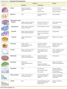
Organelles are specialized structures within the cytoplasm that perform specific functions. They are classified as membranous (e.g., mitochondria, endoplasmic reticulum) or nonmembranous (e.g., ribosomes, cytoskeleton).
Membranous: Peroxisomes, lysosomes, endoplasmic reticulum, Golgi apparatus
Nonmembranous: Cytoskeleton, centrioles, ribosomes
Cell Cycle and Division
Phases of the Cell Cycle
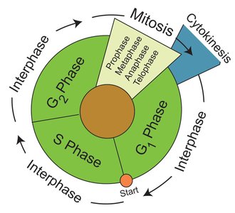
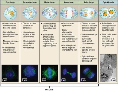
All cells undergo a life cycle consisting of interphase (growth and DNA replication) and mitotic phase (division). Interphase includes G1 (growth), S (DNA synthesis), and G2 (growth). Mitosis is the process by which somatic cells divide, producing two identical daughter cells.
Interphase: Cell grows and replicates DNA.
Mitosis: Division of nucleus (prophase, metaphase, anaphase, telophase).
Cytokinesis: Division of cytoplasm.
Plasma Membrane Structure and Function
Fluid Mosaic Model and Membrane Components
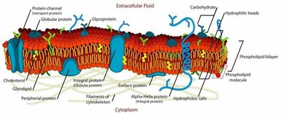
The plasma membrane is a selectively permeable barrier composed of a phospholipid bilayer, proteins, cholesterol, and carbohydrates. It separates the cell's interior from the external environment and facilitates communication and transport.
Phospholipid Bilayer: Hydrophilic heads face outward, hydrophobic tails inward.
Proteins: Integral (transmembrane) and peripheral; functions include transport, recognition, enzymatic activity, and cell joining.
Glycocalyx: Sugar coating for cell recognition.
Cholesterol: Stabilizes membrane fluidity.
Transport Across the Plasma Membrane
Passive and Active Processes
Substances move across the plasma membrane via passive (no energy required) or active (energy required) processes. Passive transport includes diffusion and osmosis; active transport uses ATP to move substances against concentration gradients.
Passive Transport: Movement from high to low concentration (diffusion, osmosis).
Active Transport: Movement from low to high concentration, requires ATP and carrier proteins.
Vesicular Transport: Endocytosis (into cell), exocytosis (out of cell), phagocytosis (cell eating), pinocytosis (cell drinking), receptor-mediated endocytosis.
Osmosis and Tonicity
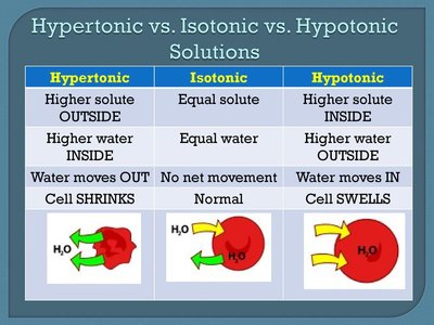
Osmosis is the movement of water across a membrane. Tonicity describes the effect of a solution on cell volume: hypertonic (cell shrinks), hypotonic (cell swells), isotonic (no change).
Type | Solute | Water | Effect on Cell |
|---|---|---|---|
Hypertonic | Higher outside | Higher inside | Cell shrinks |
Isotonic | Equal | Equal | No change |
Hypotonic | Higher inside | Higher outside | Cell swells |
Chromosome Structure and Cell Division
Chromosomes and Mitosis
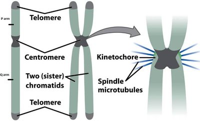
Chromosomes are composed of DNA and proteins, separated during mitosis. Each chromosome consists of two sister chromatids joined by a centromere. The kinetochore is a protein structure essential for chromosome separation.
DNA Structure and Replication
Double Helix and Base Pairing
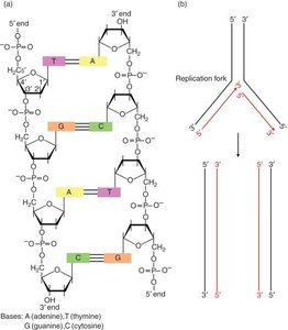
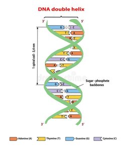
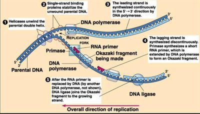
DNA is a double helix composed of nucleotides (adenine, thymine, cytosine, guanine). Replication is semiconservative, producing two identical DNA molecules. Key enzymes include helicase, primase, DNA polymerase, and ligase.
Protein Synthesis
Transcription and Translation
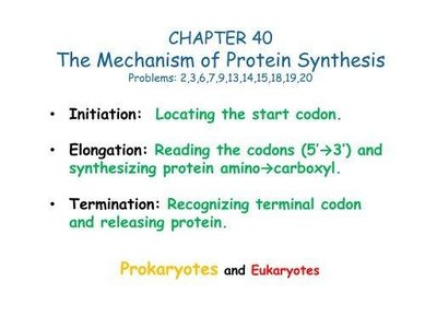
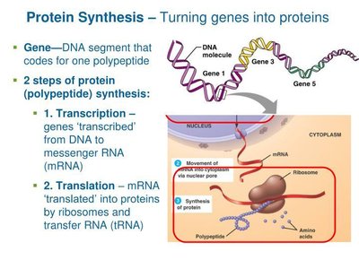
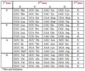
Protein synthesis involves two main steps: transcription (DNA to mRNA) and translation (mRNA to protein). Ribosomes, tRNA, and rRNA are essential for this process. Amino acids are assembled into polypeptides according to the genetic code.
Transcription: DNA makes mRNA in the nucleus.
Translation: mRNA is decoded by ribosomes and tRNA to synthesize proteins.
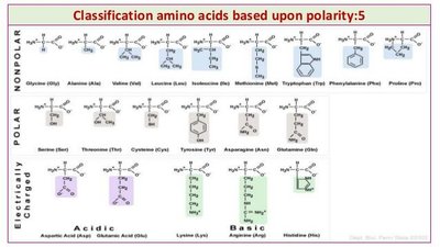
Amino Acids: 20 types, each with amine, carboxyl, and R group.
Genetic Terms and Concepts
Key Definitions
Genome: All DNA in a cell's nucleus.
Gene: Segment of DNA coding for a trait.
Exons/Introns: Coding/non-coding regions of DNA.
Karyotype: Chart of chromosomes.
Allele: Different forms of a gene.
Mutation: Permanent change in DNA sequence.
Genotype: Genetic makeup.
Phenotype: Physical expression.
Summary
Cell structure and function
Transport mechanisms across membranes
Cell cycle and division
DNA structure and replication
Protein synthesis
Genetic terminology
