 Back
BackChapter 11: Fundamentals of the Nervous System and Nervous Tissue
Study Guide - Smart Notes
 Tailored notes based on your materials, expanded with key definitions, examples, and context.
The Nervous System: Overview and Functions
Introduction to the Nervous System
The nervous system is the primary control and communication network of the body, coordinating rapid and specific responses to internal and external stimuli. It achieves this through the generation and transmission of electrical and chemical signals known as nerve impulses or action potentials.
Communication: Electrical and chemical signals allow for immediate responses.
Functions: Sensory input, integration, and motor output.
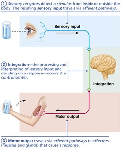
Three Main Functions of the Nervous System
Sensory Input: Sensory receptors detect changes inside or outside the body and send information to the central nervous system (CNS).
Integration: The CNS processes and interprets sensory input, deciding on an appropriate response.
Motor Output: The CNS sends signals to effectors (muscles or glands) to produce a response.
Organization of the Nervous System
Anatomical Divisions
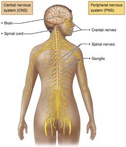
The nervous system is divided into two main parts:
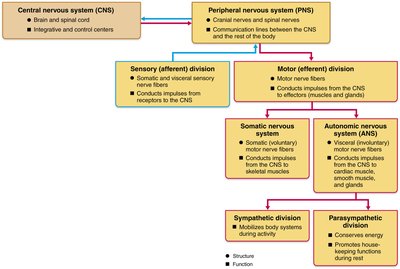
Central Nervous System (CNS): Consists of the brain and spinal cord. It is the integration and control center.
Peripheral Nervous System (PNS): Includes all neural tissue outside the CNS, mainly nerves extending from the brain and spinal cord (cranial and spinal nerves), and ganglia.
Functional Divisions of the PNS
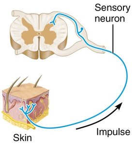
Sensory (Afferent) Division: Transmits sensory information from receptors to the CNS.
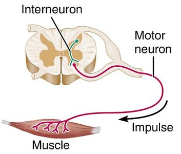
Motor (Efferent) Division: Transmits commands from the CNS to effectors (muscles and glands).
Somatic Nervous System: Controls voluntary movements of skeletal muscles.
Autonomic Nervous System (ANS): Regulates involuntary functions (smooth muscle, cardiac muscle, glands).
Sympathetic Division: Mobilizes body systems during activity (fight or flight).
Parasympathetic Division: Conserves energy and promotes housekeeping functions during rest.
Nervous Tissue: Cell Types and Structure
Principal Cell Types
Neurons (Nerve Cells): Excitable cells that transmit electrical signals.
Neuroglia (Glial Cells): Support, protect, and insulate neurons. Types include astrocytes, oligodendrocytes (CNS), and Schwann cells (PNS).
Neuroglia in the CNS and PNS
Astrocytes (CNS): Support neurons, regulate the chemical environment, and recycle neurotransmitters.
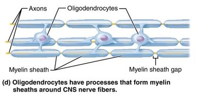
Oligodendrocytes (CNS): Form myelin sheaths around CNS axons, increasing conduction speed.
Schwann Cells (PNS): Form myelin sheaths around PNS axons and aid in nerve regeneration.
Neurons: Structure and Classification
Basic Structure of a Neuron
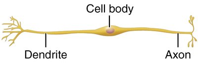
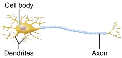
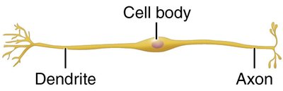
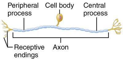
Cell Body (Soma): Contains the nucleus and organelles; site of metabolic activity.
Dendrites: Short, branched processes that receive signals from other neurons.
Axon: Long process that transmits impulses away from the cell body to other neurons or effectors.
Myelin Sheath
Myelin Sheath: Fatty, insulating layer around axons that increases the speed of impulse transmission.
Nodes of Ranvier: Gaps in the myelin sheath where action potentials are regenerated.
Myelination in CNS: Oligodendrocytes wrap multiple axons.
Myelination in PNS: Schwann cells wrap individual axons.
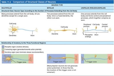
Structural Classification of Neurons
Multipolar Neurons: One axon and two or more dendrites; most common type in CNS.
Bipolar Neurons: One axon and one dendrite; found in special sensory organs (e.g., retina).
Unipolar (Pseudounipolar) Neurons: Single process that splits into peripheral and central processes; common in sensory neurons of the PNS.
Comparison of Structural Classes of Neurons
Neuron Type | Structure | Location/Function |
|---|---|---|
Multipolar | Many dendrites, one axon | Most CNS neurons, motor neurons |
Bipolar | One dendrite, one axon | Special senses (retina, olfactory epithelium) |
Unipolar | One process splits into two branches | Sensory neurons in PNS |
Functional Classification of Neurons
Sensory (Afferent) Neurons: Transmit impulses from sensory receptors toward the CNS; mostly unipolar.
Motor (Efferent) Neurons: Carry impulses from the CNS to effectors; mostly multipolar.
Interneurons (Association Neurons): Connect sensory and motor neurons within the CNS; mostly multipolar.
Membrane Potentials and Ion Channels
Resting Membrane Potential (Vm)
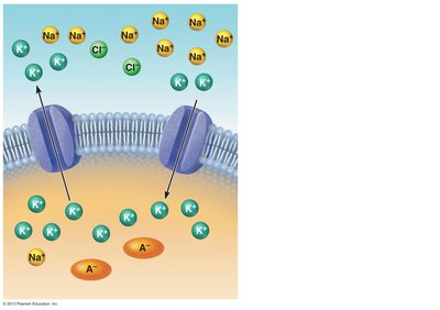
Neurons maintain a resting membrane potential (typically –70 mV) due to differences in ion concentrations and membrane permeability. This potential is essential for the generation of action potentials.
Key Factors: Sodium-potassium pump, differential permeability to K+ and Na+, presence of negatively charged proteins inside the cell.
Types of Ion Channels
Leak (Non-gated) Channels: Always open; responsible for resting Vm.
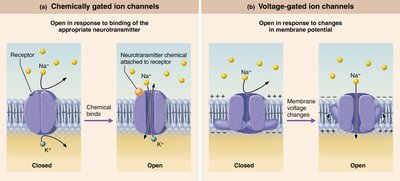
Chemically Gated (Ligand-Gated) Channels: Open in response to neurotransmitter binding; found in receptive regions.
Voltage-Gated Channels: Open in response to changes in membrane potential; found in conducting and secretory regions.
Mechanically Gated Channels: Open in response to physical deformation; found in sensory receptors.
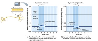
Changes in Membrane Potential
Depolarization: Membrane potential becomes less negative (e.g., –70 mV to –55 mV); increases likelihood of generating an impulse.
Hyperpolarization: Membrane potential becomes more negative (e.g., –70 mV to –90 mV); decreases likelihood of generating an impulse.
Summary Table: Factors Contributing to Resting Vm
Factor | Description |
|---|---|
Na+/K+ Pump | Maintains ion gradients by pumping 3 Na+ out and 2 K+ in |
Membrane Permeability | More permeable to K+ than Na+, leading to net negative charge inside |
Leak Channels | K+ leaks out more readily than Na+ leaks in |
Negatively Charged Proteins | Remain inside the cell, contributing to negative charge |
Location of Gated Channels in Neurons
Chemically-Gated Channels: Located in the receptive region (dendrites and cell body).
Voltage-Gated Na+ and K+ Channels: Located in the conducting region (axon).
Voltage-Gated Ca2+ Channels: Located in the secretory region (axon terminals).
Summary
The nervous system is essential for sensory input, integration, and motor output.
It is organized into CNS and PNS, with further functional subdivisions.
Neurons are classified structurally and functionally, with specialized support from neuroglia.
Membrane potentials and ion channels underlie the electrical activity of neurons.
