 Back
BackChapter 12: The Nervous System and Nervous Tissue – Study Notes
Study Guide - Smart Notes
 Tailored notes based on your materials, expanded with key definitions, examples, and context.
The Nervous System and Nervous Tissue
Overview of the Nervous System
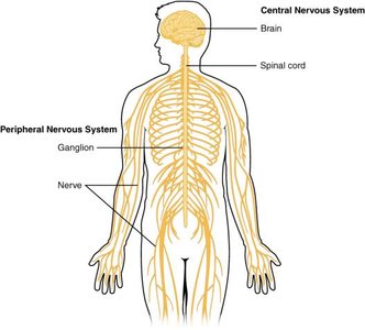
The nervous system is a complex network responsible for coordinating the body's activities by transmitting signals to and from different parts. It is divided into two main regions: the Central Nervous System (CNS) and the Peripheral Nervous System (PNS).
Central Nervous System (CNS): Consists of the brain and spinal cord. It is the main control center for processing information and directing responses.
Peripheral Nervous System (PNS): Includes all neural tissue outside the CNS, such as nerves and ganglia. It connects the CNS to limbs and organs.

Types of Cells in Nervous Tissue
Nervous tissue is composed of two primary cell types: neurons and glial cells.
Neurons: The communicative cells responsible for transmitting electrical signals throughout the nervous system.
Glial Cells: Provide structural and metabolic support to neurons, maintain homeostasis, and form myelin.
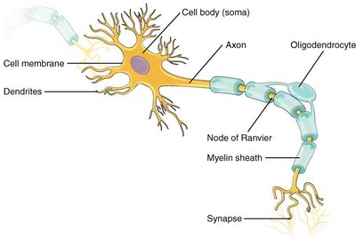
Anatomy of a Neuron
Neurons have specialized structures that enable them to receive, process, and transmit information:
Cell Body (Soma): Contains the nucleus and most organelles.
Dendrites: Extensions that receive signals from other neurons.
Axon: A single, long projection that transmits signals away from the cell body. Axons may branch to communicate with multiple target cells.
Myelin Sheath: An insulating layer produced by glial cells (oligodendrocytes in the CNS, Schwann cells in the PNS) that increases the speed of signal transmission.
Nodes of Ranvier: Gaps in the myelin sheath that facilitate rapid signal conduction (saltatory conduction).
Axon Terminal and Synapse: The axon ends in terminals that form synapses with target cells, allowing communication via neurotransmitters.
Organization of Nervous Tissue
Nervous tissue is organized into distinct structures in the CNS and PNS:
Nucleus (CNS): A localized collection of neuron cell bodies within the CNS.
Ganglion (PNS): A localized collection of neuron cell bodies in the PNS.
Tract (CNS): A bundle of axons within the CNS.
Nerve (PNS): A bundle of axons in the PNS.

Functional Divisions of the Nervous System
The nervous system performs three basic functions: sensation, integration, and response.
Sensation: Sensory receptors detect changes (stimuli) in the internal or external environment and send information to the CNS via sensory (afferent) pathways.
Integration: The CNS processes and interprets sensory input, integrating it with past experiences and current conditions to determine an appropriate response.
Response: The nervous system initiates a response through motor (efferent) pathways, which may be voluntary (somatic) or involuntary (autonomic).

Divisions of the Peripheral Nervous System
The PNS is further divided based on function:
Somatic Nervous System (SNS): Controls voluntary movements by innervating skeletal muscles.
Autonomic Nervous System (ANS): Regulates involuntary functions such as heart rate, digestion, and glandular activity. It is subdivided into the sympathetic and parasympathetic divisions.
Enteric Nervous System (ENS): Sometimes considered part of the ANS, it independently controls the digestive tract.
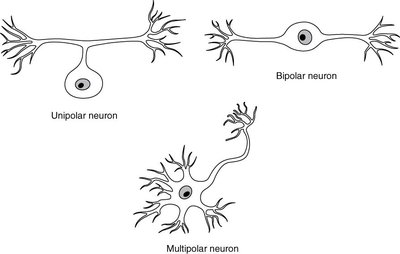
Types of Neurons
Neurons are classified based on the number and arrangement of their processes:
Type | Structure | Location/Function |
|---|---|---|
Unipolar | Single process (axon) | Found in invertebrates; humans have pseudo-unipolar neurons in sensory pathways |
Bipolar | One axon, one dendrite | Rare; found in olfactory epithelium and retina |
Multipolar | One axon, two or more dendrites | Most common; motor and interneurons |
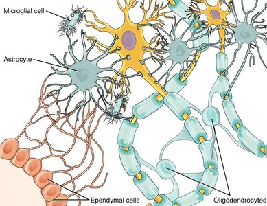
Glial Cells (Neuroglia)
Glial cells support and protect neurons. They differ between the CNS and PNS:
CNS Glial Cells:
Astrocytes: Regulate extracellular ion concentration, form the blood-brain barrier, and remove excess neurotransmitters.
Oligodendrocytes: Myelinate axons in the CNS.
Ependymal Cells: Produce and circulate cerebrospinal fluid (CSF).
Microglia: Act as immune cells within the CNS.
PNS Glial Cells:
Satellite Cells: Support neuron cell bodies in ganglia.
Schwann Cells: Myelinate axons in the PNS (each Schwann cell myelinates a single axon segment).
Ion Channels and Membrane Potential
The cell membrane separates the intracellular and extracellular environments, and ions move across the membrane through specialized proteins called ion channels. The distribution of ions creates a membrane potential (measured in millivolts, mV).
Ligand-Gated Channels: Open in response to binding of a chemical messenger (ligand), such as a neurotransmitter.
Mechanically Gated Channels: Open in response to physical deformation of the membrane.
Voltage-Gated Channels: Open in response to changes in membrane potential.
At rest, the inside of the neuron is negatively charged relative to the outside (resting membrane potential ≈ -70 mV). This is maintained by the sodium-potassium pump and ion leakage channels.
Action Potentials
An action potential is a rapid, temporary change in membrane potential that allows neurons to transmit signals over long distances. It is an all-or-none event:
A stimulus depolarizes the membrane to threshold (-55 mV).
Voltage-gated sodium channels open, sodium enters, and the cell rapidly depolarizes to +30 mV.
At +30 mV, sodium channels close and voltage-gated potassium channels open, repolarizing the cell.
Potassium channels close slowly, causing a brief hyperpolarization before returning to resting potential.
Graded potentials are local changes in membrane potential that can trigger action potentials if they reach threshold.
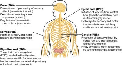
Summary Table: Key Structures and Functions
Structure | Location | Function |
|---|---|---|
Brain (CNS) | Cranial cavity | Processing sensory input, initiating motor output, regulating homeostasis |
Spinal Cord (CNS) | Vertebral canal | Conducting signals to/from brain, reflexes |
Nerves (PNS) | Throughout body | Transmit sensory and motor signals |
Ganglia (PNS) | Near spinal cord and organs | Relay sensory and autonomic signals |
Digestive Tract (ENS) | Digestive organs | Autonomous control of digestion |
