Back
BackChapter 17: Blood – Structure, Function, and Clinical Relevance
Study Guide - Smart Notes
 Tailored notes based on your materials, expanded with key definitions, examples, and context.
Blood: Overview and Functions
Introduction to Blood
Blood is a specialized connective tissue that plays a critical role in the transport of substances, regulation of physiological parameters, and protection against disease. It is the only fluid tissue in the human body and is essential for maintaining homeostasis.
Transport: Delivers oxygen and nutrients, removes metabolic wastes, and transports hormones.
Regulation: Maintains body temperature, pH balance, and fluid volume.
Protection: Prevents blood loss through clotting and combats infection via immune cells and proteins.
Composition of Blood
Major Components
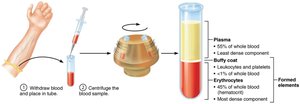
Blood consists of a liquid matrix called plasma and formed elements (cells and cell fragments) suspended within it. When centrifuged, blood separates into three layers:
Plasma: ~55% of whole blood; the least dense component.
Buffy coat: <1% of blood; contains leukocytes (white blood cells) and platelets.
Erythrocytes: ~45% of blood (hematocrit); the most dense component.
Blood Plasma
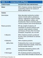
Plasma is a straw-colored, sticky fluid that makes up about 90% water and contains over 100 dissolved solutes, including nutrients, gases, hormones, wastes, proteins, and inorganic ions. Plasma proteins, mainly produced by the liver, are the most abundant solutes and serve various functions such as transport, buffering, and maintaining osmotic pressure.
Constituent | Description and Importance |
|---|---|
Water | 90% of plasma volume; dissolving and suspending medium for solutes of blood; absorbs heat |
Electrolytes | Most abundant solutes by number; help maintain plasma osmotic pressure and normal blood pH |
Plasma proteins | Albumin (60%): carrier, buffer, osmotic pressure; Globulins (36%): transport and antibodies; Fibrinogen (4%): forms fibrin threads of blood clot |
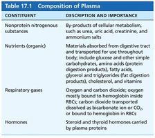
Constituent | Description and Importance |
|---|---|
Nonprotein nitrogenous substances | By-products of metabolism (urea, uric acid, creatinine, etc.) |
Nutrients (organic) | Materials absorbed from digestive tract (glucose, amino acids, fatty acids, etc.) |
Respiratory gases | Oxygen (bound to hemoglobin) and carbon dioxide (dissolved or as bicarbonate) |
Hormones | Steroid and thyroid hormones carried by plasma proteins |
Formed Elements
The formed elements of blood include erythrocytes (red blood cells), leukocytes (white blood cells), and platelets. Only leukocytes are complete cells; erythrocytes lack nuclei and organelles, and platelets are cell fragments. Most formed elements are short-lived and originate in the bone marrow.
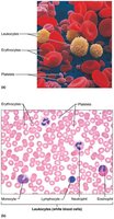
Erythrocytes (Red Blood Cells)
Structure and Function
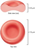
Erythrocytes are small, biconcave, anucleate cells specialized for gas transport. Their shape increases surface area for gas exchange, and they are filled with hemoglobin, the protein responsible for oxygen transport. The absence of mitochondria ensures that erythrocytes do not consume the oxygen they carry.
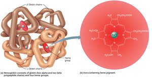
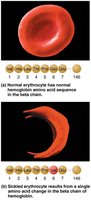
Hemoglobin: Each molecule consists of four polypeptide chains (two alpha, two beta) and four heme groups, each containing an iron atom that binds oxygen.
Oxygen transport: Each RBC contains about 250 million hemoglobin molecules, each carrying up to four oxygen molecules.
Production and Regulation
Red blood cell production (erythropoiesis) occurs in red bone marrow from hematopoietic stem cells. The process is regulated by the hormone erythropoietin (EPO), primarily released by the kidneys in response to hypoxia (low oxygen levels).
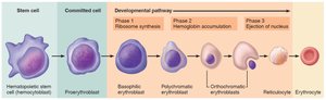
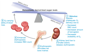
Dietary requirements: Iron, vitamin B12, folic acid, amino acids, lipids, and carbohydrates are essential for erythropoiesis.
Disorders of Erythrocytes
Anemia: Reduced oxygen-carrying capacity due to blood loss, insufficient RBC production, or increased RBC destruction. Symptoms include fatigue, pallor, and shortness of breath.
Sickle-cell anemia: Genetic disorder causing abnormal hemoglobin (HbS), leading to crescent-shaped RBCs that rupture easily and block vessels.
Leukocytes (White Blood Cells)
Types and Functions
Leukocytes are the only complete cells among the formed elements and are crucial for defense against disease. They are classified as granulocytes (with visible granules) or agranulocytes (without visible granules).
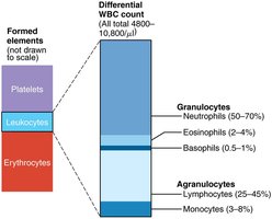
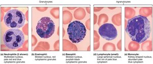
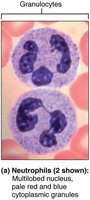
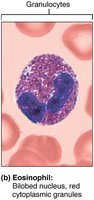
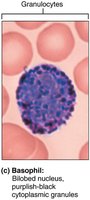
Granulocytes: Neutrophils (bacteria slayers, phagocytic), eosinophils (combat parasites, modulate allergies), basophils (release histamine in inflammation).
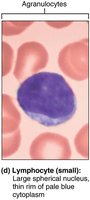
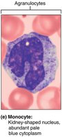
Agranulocytes: Lymphocytes (T cells and B cells, crucial for immunity), monocytes (differentiate into macrophages, phagocytic).
Leukocyte Disorders
Leukopenia: Abnormally low WBC count, often drug-induced.
Leukemias: Cancers involving overproduction of abnormal WBCs, leading to anemia, bleeding, and increased infection risk.
Platelets
Structure and Function
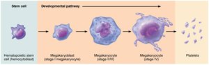
Platelets are cell fragments derived from megakaryocytes. They play a vital role in hemostasis by forming temporary plugs to seal breaks in blood vessels and releasing chemicals necessary for clotting.
Hemostasis
Steps of Hemostasis
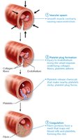
Hemostasis is the process of stopping bleeding and involves three major steps:
Vascular spasm: Vasoconstriction of damaged blood vessel.
Platelet plug formation: Platelets adhere to exposed collagen fibers and release chemicals to recruit more platelets.
Coagulation: Reinforcement of the platelet plug with fibrin threads, forming a stable blood clot.
Coagulation Pathways
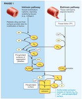
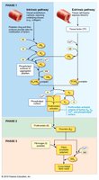
Coagulation involves a cascade of clotting factors leading to the conversion of fibrinogen to fibrin. There are two initial pathways (intrinsic and extrinsic) that converge to a common pathway.
Intrinsic pathway: Initiated by factors within the blood.
Extrinsic pathway: Initiated by external tissue damage.
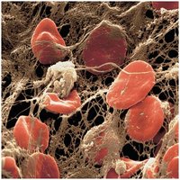
Fibrin Mesh and Clot Retraction
Thrombin converts soluble fibrinogen into insoluble fibrin, which forms a mesh that traps blood cells and seals the vessel. Clot retraction and fibrinolysis ensure the clot is removed after healing.
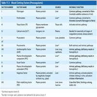
Blood Clotting Factors
Factor Number | Factor Name | Source | Primary Function |
|---|---|---|---|
I | Fibrinogen | Liver | Converted to fibrin in clot formation |
II | Prothrombin | Liver | Converted to thrombin |
IV | Calcium ions (Ca2+) | Diet, bones, plasma | Essential for all stages of coagulation |
VIII | Antihemophilic factor (AHF) | Liver, lung, capillaries | Intrinsic pathway; deficiency causes hemophilia A |
Blood Groups and Transfusions
ABO and Rh Blood Groups
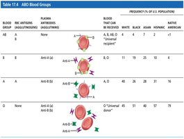
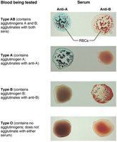
Blood groups are determined by the presence or absence of specific antigens (agglutinogens) on the surface of RBCs. The ABO system is based on A and B antigens, while the Rh system is based on the D antigen.
Blood Group | RBC Antigens | Plasma Antibodies | Blood That Can Be Received |
|---|---|---|---|
A | A | Anti-B | A, O |
B | B | Anti-A | B, O |
AB | A, B | None | A, B, AB, O (universal recipient) |
O | None | Anti-A, Anti-B | O (universal donor) |
Blood Typing
Blood typing involves mixing blood with antibodies against A and B antigens to observe agglutination. Cross-matching ensures compatibility between donor and recipient.
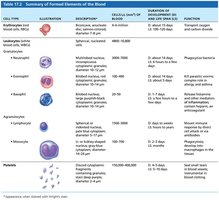
Summary Table: Formed Elements of Blood
Cell Type | Description | Number per μl | Function |
|---|---|---|---|
Erythrocyte | Biconcave, anucleate | 4–6 million | Transport O2 and CO2 |
Neutrophil | Multilobed nucleus, pale granules | 3000–7000 | Phagocytize bacteria |
Lymphocyte | Large nucleus, thin rim of cytoplasm | 1500–3000 | Immunity (T and B cells) |
Monocyte | Kidney-shaped nucleus | 100–700 | Phagocytosis, develop into macrophages |
Eosinophil | Bilobed nucleus, red granules | 100–400 | Kill parasitic worms, modulate allergies |
Basophil | Bilobed nucleus, purplish-black granules | 20–50 | Release histamine, inflammation |
Platelets | Cell fragments | 150,000–400,000 | Seal small tears, clotting |