 Back
BackChapter 19: Blood – Structure, Function, and Clinical Relevance
Study Guide - Smart Notes
 Tailored notes based on your materials, expanded with key definitions, examples, and context.
Blood and the Cardiovascular System
Overview of the Cardiovascular System
The cardiovascular system is essential for transporting substances throughout the body. It consists of the heart (a pump), blood vessels (conducting hoses), and blood (a fluid connective tissue). Blood plays a critical role in maintaining homeostasis and supporting cellular function.
Blood is a specialized connective tissue with cells suspended in a fluid matrix.
Functions include transporting gases, nutrients, hormones, and wastes; regulating pH and ion composition; restricting fluid loss; defending against toxins and pathogens; and stabilizing body temperature.
Components and Functions of Blood
Physical and Chemical Characteristics
Temperature: 38ºC (100.4ºF)
Viscosity: High
pH: Slightly alkaline (7.35–7.45)
Volume: ~7% of body weight (e.g., 5.25 L in a 75-kg adult)
Composition of Whole Blood
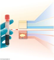
Whole blood consists of plasma (fluid) and formed elements (cells and cell fragments).
Fractionation is the process of separating whole blood into plasma and formed elements.
Plasma
Makes up about 55% of blood volume.
Over 90% is water; also contains plasma proteins and other solutes.
Similar to interstitial fluid but differs in protein composition.
Formed Elements
Comprise 37–54% of blood volume.
Include platelets, white blood cells (WBCs), and red blood cells (RBCs).
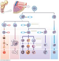
Produced by hemopoiesis (hematopoiesis) from myeloid and lymphoid stem cells.

Plasma Proteins
Albumins (60%): Maintain osmotic pressure, transport fatty acids and hormones.
Globulins (35%): Include antibodies (immunoglobulins) and transport proteins.
Fibrinogen (4%): Functions in clotting; converted to fibrin during coagulation.
Other proteins (1%): Enzymes and hormones.
Most plasma proteins are synthesized in the liver; antibodies are produced by plasma cells.
Red Blood Cells (Erythrocytes)
Structure and Function
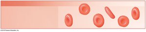
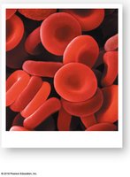
Red blood cells (RBCs) are the most abundant formed elements, specialized for oxygen and carbon dioxide transport.
Account for 99.9% of formed elements.
Contain hemoglobin, a red pigment responsible for gas transport.
Normal RBC count: Males 4.5–6.3 million/μL; Females 4.2–5.5 million/μL.
Hematocrit: Percentage of formed elements in blood (Males ~46%, Females ~42%).
Shape and Structure
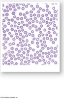
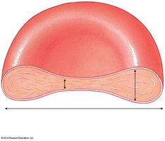
Biconcave discs: Thin central region, thicker outer margin.
Anucleate (no nucleus), lack mitochondria and ribosomes.
Cannot divide or repair; lifespan ~120 days.
Hemoglobin Structure and Function
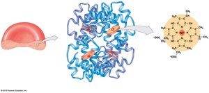
Composed of four globular protein subunits (2 alpha, 2 beta), each with a heme group containing iron.
Each iron binds one O2 molecule, forming oxyhemoglobin (HbO2).
Hemoglobin also binds CO2 (as carbaminohemoglobin) in tissues.
Normal hemoglobin: Males 14–18 g/dL; Females 12–16 g/dL.
RBC Life Cycle and Erythropoiesis
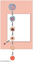
~1% of RBCs replaced daily; ~3 million new RBCs/second.
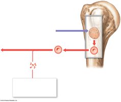
Erythropoiesis: RBC formation, occurs in red bone marrow in adults.
Regulated by erythropoietin (EPO), a hormone released by kidneys and liver in response to hypoxia.
Requires amino acids, iron, folic acid, vitamins B12 and B6.
Lack of vitamin B12 causes pernicious anemia.
Hemoglobin and Iron Recycling
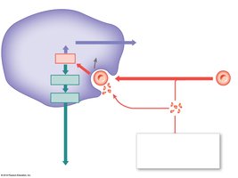
Macrophages in spleen, liver, and bone marrow engulf old RBCs and recycle hemoglobin.
Iron is removed from heme, transported by transferrin, and stored as ferritin or hemosiderin.
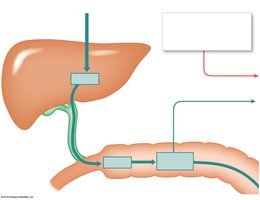
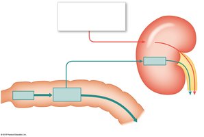
Heme is converted to biliverdin, then bilirubin (excreted in bile); excess bilirubin causes jaundice.
Breakdown products (urobilins, stercobilins) are excreted in urine and feces.
Blood Types
Surface Antigens and Blood Groups
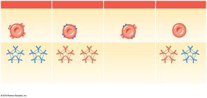
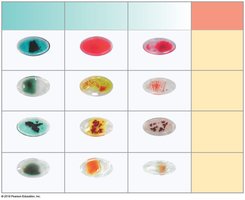
Blood type is determined by the presence or absence of surface antigens (A, B, Rh) on RBCs.
Four main blood types: A, B, AB, O.
Rh group: Rh+ (antigen present), Rh– (antigen absent).
Agglutinogens and Agglutinins
Agglutinogens: Surface antigens on RBCs.
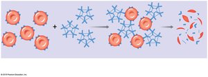
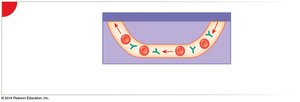
Agglutinins: Antibodies in plasma that attack foreign antigens, causing agglutination (clumping).
Type A: Anti-B antibodies; Type B: Anti-A antibodies; Type O: Both anti-A and anti-B; Type AB: Neither antibody.
Only sensitized Rh– individuals have anti-Rh antibodies.
Hemolytic Disease of the Newborn (HDN)
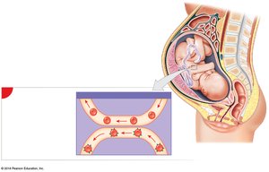
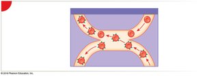
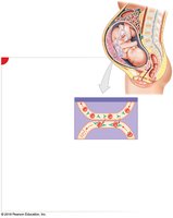
Occurs when an Rh– mother carries an Rh+ fetus.
First pregnancy usually unaffected; sensitization occurs at delivery.
Subsequent Rh+ pregnancies: Maternal anti-Rh antibodies cross placenta, destroy fetal RBCs, causing anemia and jaundice.
Prevention: Administration of RhoGAM to the mother prevents antibody formation.
White Blood Cells (Leukocytes)
General Features and Functions
Have nuclei and organelles, but lack hemoglobin.
Defend against pathogens, remove toxins and wastes, attack abnormal or damaged cells.
Types of White Blood Cells
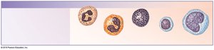
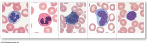
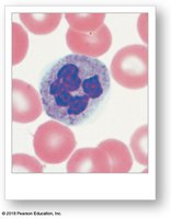
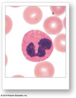
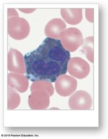
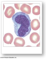
Neutrophils: 50–70% of WBCs; phagocytic, attack bacteria, contribute to pus.
Eosinophils: 2–4%; attack parasites, modulate allergic responses.
Basophils: <1%; release histamine (vasodilation) and heparin (anticoagulant).
Monocytes: 2–8%; become macrophages in tissues, engulf large pathogens.
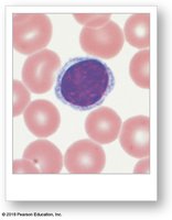
Lymphocytes: 20–40%; specific immunity, include T cells, B cells, and NK cells.
Lymphocyte Classes
T cells: Cell-mediated immunity, attack foreign cells, regulate immune response.
B cells: Humoral immunity, differentiate into plasma cells that produce antibodies.
Natural Killer (NK) cells: Destroy abnormal cells (e.g., cancerous or virus-infected).
Clinical Relevance
Differential count helps detect infection, inflammation, and allergic reactions.
Leukopenia: Low WBC count; Leukocytosis: High WBC count; Leukemia: Cancer of WBCs.
Platelets (Thrombocytes)
Structure and Function
Cell fragments involved in clotting; circulate 9–12 days.
Removed by phagocytes (mainly in spleen); 150,000–500,000/μL blood.
Stored in vascular organs, mobilized during crisis.
Functions of Platelets
Release clotting chemicals.
Temporarily patch damaged vessel walls.
Reduce size of vessel wall break.
Thrombocytopoiesis
Platelet production in red bone marrow by megakaryocytes (giant cells that shed cytoplasmic fragments).
Hemostasis
Phases of Hemostasis
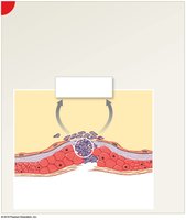
Hemostasis is the process of stopping bleeding and involves three phases:
Vascular phase: Endothelial cells contract, release endothelins, and become sticky to reduce blood flow.
Platelet phase: Platelets adhere to damaged area, aggregate, and release chemicals to promote clotting and repair.
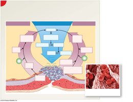
Coagulation phase: Cascade of reactions converts fibrinogen to fibrin, forming a stable blood clot.
Clot Retraction
After clot formation, platelets contract, pulling torn edges together and reducing the size of the damaged area.
Summary Table: Main Components of Blood
Component | Percentage | Main Function |
|---|---|---|
Plasma | ~55% | Transport of nutrients, hormones, proteins, and wastes |
Red Blood Cells | ~45% | Oxygen and carbon dioxide transport |
White Blood Cells | <1% | Defense against pathogens |
Platelets | <1% | Clotting |
Additional info: This guide covers the essential structure and function of blood, its components, and clinical relevance, as outlined in a typical Anatomy & Physiology curriculum.
