Back
BackEndocrine, Blood, Heart, and Blood Vessel Systems: Structured Study Notes
Study Guide - Smart Notes
 Tailored notes based on your materials, expanded with key definitions, examples, and context.
Endocrine System
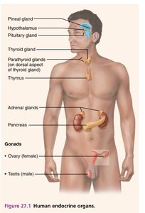
Overview of Human Endocrine Organs
The endocrine system consists of glands that secrete hormones directly into the bloodstream, regulating various physiological processes. Major endocrine organs include the pineal gland, hypothalamus, pituitary gland, thyroid gland, parathyroid glands, thymus, adrenal glands, pancreas, and gonads (ovaries and testes).
Pineal gland: Produces melatonin, regulating sleep cycles.
Hypothalamus: Controls pituitary gland and links nervous and endocrine systems.
Pituitary gland: Master gland, regulates other endocrine glands.
Thyroid gland: Controls metabolism.
Parathyroid glands: Regulate calcium levels.
Thymus: Involved in immune function.
Adrenal glands: Produce hormones for stress response and metabolism.
Pancreas: Regulates blood glucose.
Gonads: Produce sex hormones.
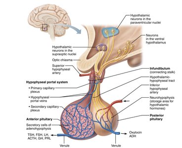
Hypothalamic-Pituitary Axis
The hypothalamus and pituitary gland form a central regulatory axis for endocrine function. The hypothalamus produces releasing and inhibiting hormones that control the anterior pituitary, while the posterior pituitary stores and releases hormones produced by the hypothalamus.
Anterior pituitary: Secretes hormones such as TSH, FSH, LH, ACTH, GH, and PRL.
Posterior pituitary: Stores and releases oxytocin and ADH.
Infundibulum: Connects hypothalamus to pituitary.
Hypophyseal portal system: Transports hormones from hypothalamus to anterior pituitary.
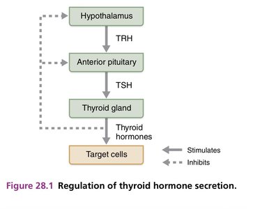
Regulation of Thyroid Hormone Secretion
Thyroid hormone secretion is regulated by a feedback loop involving the hypothalamus, anterior pituitary, and thyroid gland. The hypothalamus releases TRH, stimulating the anterior pituitary to release TSH, which then stimulates the thyroid gland to produce thyroid hormones. These hormones act on target cells and inhibit further release of TRH and TSH.
TRH: Thyrotropin-releasing hormone from hypothalamus.
TSH: Thyroid-stimulating hormone from anterior pituitary.
Thyroid hormones: Regulate metabolism and inhibit TRH/TSH release.
Summary of Pituitary Gland Hormones
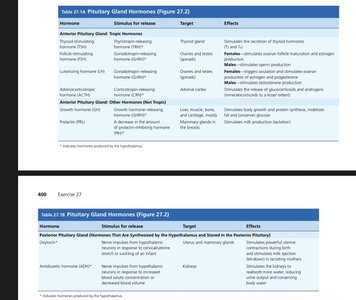
The pituitary gland produces several hormones, each with specific targets and effects. The anterior pituitary releases tropic hormones that regulate other glands, while the posterior pituitary stores hormones produced by the hypothalamus.
Hormone | Stimulus for Release | Target | Effects |
|---|---|---|---|
TSH | TRH | Thyroid gland | Stimulates thyroid hormone secretion |
FSH | GnRH | Ovaries/testes | Stimulates gamete production |
LH | GnRH | Ovaries/testes | Stimulates hormone production |
ACTH | CRH | Adrenal cortex | Stimulates cortisol secretion |
GH | GHRH | Body cells | Stimulates growth |
PRL | PRH | Mammary glands | Stimulates milk production |
Oxytocin | Nerve impulses | Uterus/mammary glands | Stimulates contractions and milk ejection |
ADH | Nerve impulses | Kidneys | Promotes water reabsorption |
Adrenal Gland, Pancreas, and Gonad Hormones
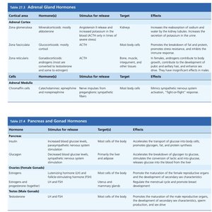
The adrenal glands, pancreas, and gonads produce hormones essential for metabolism, stress response, and reproduction. The adrenal cortex produces corticosteroids, the medulla produces catecholamines, the pancreas regulates glucose, and the gonads produce sex hormones.
Hormone | Stimulus for Release | Target | Effects |
|---|---|---|---|
Aldosterone | Low blood volume/pressure | Kidneys | Increases sodium reabsorption |
Cortisol | ACTH | Body cells | Stress response, metabolism |
Epinephrine | Sympathetic stimulation | Body cells | Fight-or-flight response |
Insulin | High blood glucose | Body cells | Lowers blood glucose |
Glucagon | Low blood glucose | Liver | Raises blood glucose |
Estrogen/Progesterone | FSH/LH | Body cells | Female reproductive function |
Testosterone | LH | Body cells | Male reproductive function |
Endocrine Homeostatic Imbalances
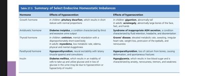
Imbalances in hormone secretion can lead to various disorders. Hyposecretion and hypersecretion of hormones have distinct effects on the body.
Hormone | Effects of Hyposecretion | Effects of Hypersecretion |
|---|---|---|
Growth hormone | Pituitary dwarfism | Gigantism, acromegaly |
ADH | Diabetes insipidus | Syndrome of inappropriate ADH secretion |
Thyroid hormone | Hypothyroidism, myxedema | Graves' disease |
Parathyroid hormone | Hypoparathyroidism | Hyperparathyroidism |
Insulin | Diabetes mellitus | Hypoglycemia |
Blood
Composition of Blood
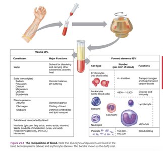
Blood is composed of plasma and formed elements. Plasma contains water, proteins, and solutes, while formed elements include erythrocytes, leukocytes, and platelets.
Plasma: 55% of blood, contains water, proteins, electrolytes, nutrients, and waste products.
Formed elements: 45% of blood, includes red blood cells (RBCs), white blood cells (WBCs), and platelets.
Formed Elements of Blood
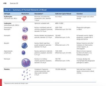
The formed elements of blood include erythrocytes, leukocytes, and platelets, each with distinct functions and characteristics.
Cell Type | Description | Cells/mm3 | Function |
|---|---|---|---|
Erythrocytes | Biconcave, anucleate | 4-6 million | Transport oxygen and carbon dioxide |
Neutrophils | Multilobed nucleus | 4800-10800 | Phagocytosis of pathogens |
Eosinophils | Bilobed nucleus | 100-400 | Kill parasitic worms |
Basophils | Large nucleus | 20-50 | Release histamine |
Lymphocytes | Small, spherical nucleus | 1500-3000 | Immune response |
Monocytes | U-shaped nucleus | 100-700 | Phagocytosis |
Platelets | Cell fragments | 150,000-400,000 | Blood clotting |
Blood Coagulation
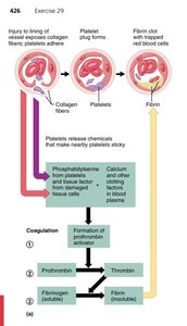
Blood coagulation is a complex process involving platelets and clotting factors. Injury exposes collagen fibers, platelets adhere, and a cascade leads to the formation of a fibrin clot.
Platelet plug formation: Platelets adhere to exposed collagen.
Coagulation cascade: Formation of prothrombin activator, conversion of prothrombin to thrombin, and fibrinogen to fibrin.
ABO Blood Typing
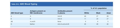
ABO blood typing is based on the presence of antigens on RBC membranes and antibodies in plasma. Blood type determines compatibility for transfusions.
ABO Blood Type | Antigens on RBC | Antibodies in Plasma | % of U.S. Population |
|---|---|---|---|
A | A | Anti-B | White: 40, Black: 27, Asian: 28 |
B | B | Anti-A | White: 11, Black: 20, Asian: 27 |
AB | A and B | None | White: 4, Black: 4, Asian: 5 |
O | Neither | Anti-A and Anti-B | White: 45, Black: 49, Asian: 40 |
Heart
Gross Anatomy of the Heart
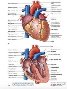
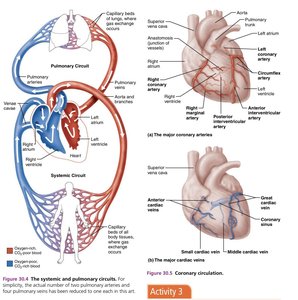
The heart is a muscular organ with four chambers: two atria and two ventricles. It contains valves that ensure unidirectional blood flow and is supplied by coronary arteries.
Chambers: Right and left atria, right and left ventricles.
Valves: Tricuspid, bicuspid (mitral), pulmonary, and aortic valves.
Coronary circulation: Supplies heart muscle with blood.
Systemic and Pulmonary Circuits
The heart pumps blood through two circuits: the pulmonary circuit (to the lungs) and the systemic circuit (to the body). Oxygen-poor blood is sent to the lungs, while oxygen-rich blood is distributed to tissues.
Pulmonary circuit: Right ventricle → lungs → left atrium.
Systemic circuit: Left ventricle → body → right atrium.
Intrinsic Conduction System of the Heart
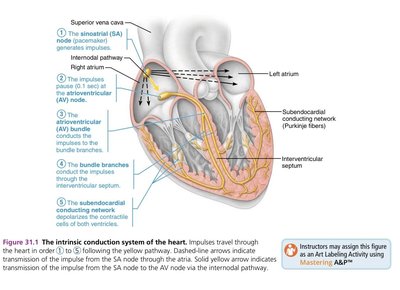
The heart's intrinsic conduction system coordinates contraction. Key components include the SA node, AV node, bundle branches, and Purkinje fibers.
SA node: Pacemaker, initiates impulse.
AV node: Delays impulse.
Bundle branches: Conduct impulse to ventricles.
Purkinje fibers: Stimulate ventricular contraction.
Electrocardiogram (ECG) and Cardiac Cycle
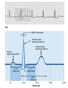
An ECG records the electrical activity of the heart. The cardiac cycle includes atrial depolarization (P wave), ventricular depolarization (QRS complex), and ventricular repolarization (T wave).
P wave: Atrial depolarization.
QRS complex: Ventricular depolarization.
T wave: Ventricular repolarization.
Blood Vessels
Generalized Structure of Arteries, Veins, and Capillaries
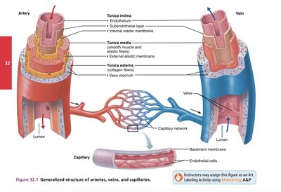
Blood vessels are classified as arteries, veins, and capillaries. Arteries have thick walls and carry blood away from the heart, veins have thinner walls and valves, and capillaries are sites of exchange.
Arteries: Tunica intima, media, and externa.
Veins: Tunica intima, media, externa, and valves.
Capillaries: Endothelial cells, basement membrane.
Systemic Arterial Circulation
The systemic arterial circulation distributes oxygenated blood throughout the body. The aorta branches into major arteries supplying the head, neck, thorax, abdomen, and limbs.
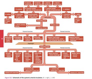
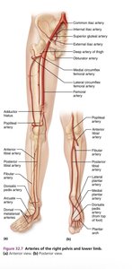
Major Arteries of the Head, Neck, Upper Limb, Abdomen, and Lower Limb
Major arteries supply specific regions of the body. The carotid arteries supply the head and neck, the subclavian and brachial arteries supply the upper limb, the abdominal aorta supplies abdominal organs, and the femoral and popliteal arteries supply the lower limb.
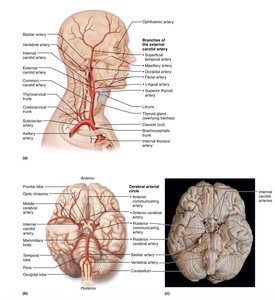
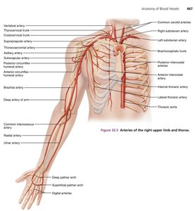
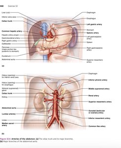
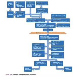
Systemic Venous Circulation
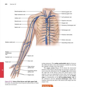
Venous circulation returns deoxygenated blood to the heart. Major veins include the jugular, subclavian, superior and inferior vena cava, and veins of the limbs and abdomen.
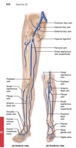
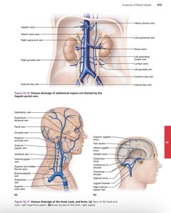
Major Veins of the Head, Neck, Upper Limb, Abdomen, and Lower Limb
Major veins drain blood from specific regions. The jugular veins drain the head and neck, the cephalic and basilic veins drain the upper limb, the hepatic portal vein drains abdominal organs, and the femoral and saphenous veins drain the lower limb.
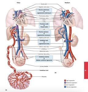
Fetal and Newborn Circulation
Fetal circulation includes unique structures such as the ductus arteriosus and foramen ovale, which bypass the lungs. After birth, these structures close, and normal circulation is established.