 Back
BackHistology and Organization of Nervous Tissue: Neurons and Neuroglia
Study Guide - Smart Notes
 Tailored notes based on your materials, expanded with key definitions, examples, and context.
Histology of Nervous Tissue
Overview of Nervous Tissue
Nervous tissue is specialized for communication and control within the body. It consists of two main cell types: neurons, which transmit electrical signals, and neuroglia (glial cells), which support and protect neurons.
Neurons: Excitable cells responsible for transmitting impulses.
Neuroglia: Non-excitable cells that provide structural, metabolic, and functional support to neurons.
Neurons
General Structure of a Neuron
Neurons have a distinct morphology that enables their function in signal transmission.
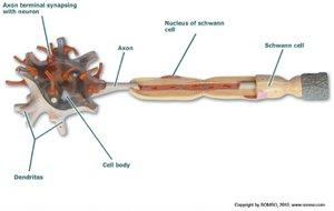
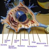
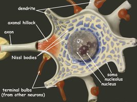
Cell Body (Soma): Contains the nucleus and Nissl bodies (rough ER for protein synthesis).
Dendrites: Branching processes that receive signals from other cells.
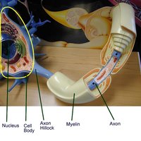
Axon: Long process that transmits impulses away from the cell body; originates at the axon hillock.
Nucleus: Central organelle containing genetic material.
Nucleolus: Site of ribosomal RNA synthesis within the nucleus.
Example: Motor neurons in the spinal cord exhibit these structures clearly. 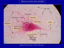
Classification of Neurons
Neurons are classified by both structure and function.
Structural Classification:
Unipolar: Single process; typical of sensory neurons.
Bipolar: Two processes; found in special sensory organs.
Multipolar: Multiple processes; most common, including motor and interneurons.
Functional Classification:
Sensory (Afferent): Transmit impulses toward CNS.
Motor (Efferent): Carry instructions from CNS to effectors.
Interneurons: Integrate information within CNS.
Example: Multipolar neurons are predominant in the CNS, while unipolar neurons are typical in sensory ganglia.
Neuroglia (Glial Cells)
Types of Neuroglia
Neuroglia are essential for nervous system function and are classified based on their location and role.
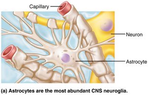
Astrocytes: Maintain blood-brain barrier, provide structural support, regulate ion and nutrient concentrations, absorb/recycle neurotransmitters, and form scar tissue after injury.
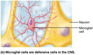
Microglia: Defensive cells; phagocytize pathogens and debris.
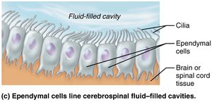
Ependymal Cells: Line cerebrospinal fluid-filled cavities; produce and circulate CSF.
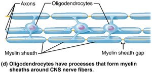
Oligodendrocytes: Form myelin sheaths in CNS.
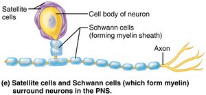
Schwann Cells: Form myelin sheaths in PNS; aid in regeneration.
Satellite Cells: Surround neuron cell bodies in PNS; regulate environment.
Myelination and Schwann Cells
Peripheral Myelination
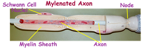
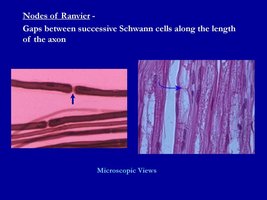
Schwann Cells: Wrap around axons in the PNS, forming the myelin sheath and neurilemma.
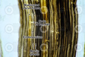
Nodes of Ranvier: Gaps between Schwann cells; facilitate rapid impulse conduction via saltatory conduction.
Central Myelination: Oligodendrocytes form myelin in the CNS.
Structure of a Nerve
Connective Tissue Coverings
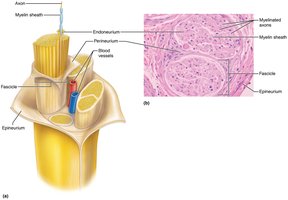
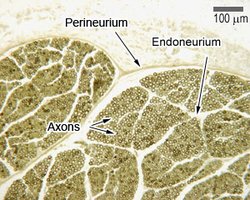
Nerves are organized into bundles and protected by connective tissue layers.
Endoneurium: Surrounds individual axons.
Perineurium: Surrounds fascicles (bundles of axons).
Epineurium: Surrounds the entire nerve.
Synapse
General Structure and Function
A synapse is the junction where a neuron communicates with another cell.
Presynaptic End Bulb: Contains neurotransmitter vesicles.
Synaptic Cleft: Gap between presynaptic and postsynaptic cells.
Postsynaptic Membrane: Contains receptors for neurotransmitters.
Mechanism: An action potential reaches the presynaptic bulb, triggering neurotransmitter release into the synaptic cleft. Neurotransmitters bind to receptors on the postsynaptic membrane, altering its potential and possibly generating a new action potential or initiating other responses. 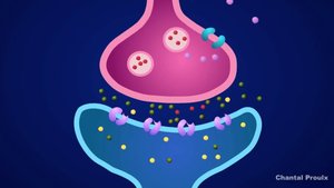
Functional Organization of the Nervous System
CNS vs. PNS
Central Nervous System (CNS): Brain and spinal cord; integration and processing center.
Peripheral Nervous System (PNS): All neural tissue outside CNS; connects CNS to the rest of the body.
Divisions of the PNS
Sensory (Afferent) Division: Transmits sensory information to CNS.
Motor (Efferent) Division: Transmits motor commands from CNS to effectors.
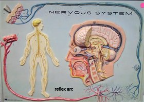
Reflex Arc
Components of a Reflex Arc
A reflex arc is the functional unit of the nervous system, enabling rapid, automatic responses to stimuli.
Receptor: Detects stimulus.
Sensory (Afferent) Neuron: Transmits impulse to CNS.
Interneuron: Integrates information within CNS.
Motor (Efferent) Neuron: Transmits impulse to effector.
Effector: Muscle or gland responding to stimulus.
Histological Examples
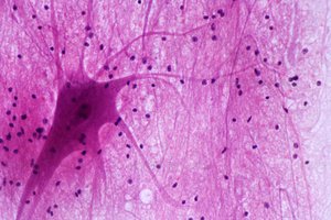
Multipolar Motor Neurons in Spinal Cord
Multipolar neurons are prominent in the spinal cord.
Neuroglia nuclei are visible as small dots in histological sections.
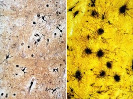
Microglia and Astrocytes in CNS
Microglia and astrocytes are two key types of neuroglia in the CNS.
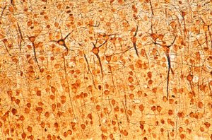
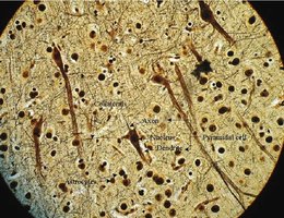
Pyramidal Cells in Cerebrum
Pyramidal cells are multipolar neurons found in the cerebral cortex.
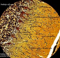
Cerebral Cortex and Cerebellum
The cerebral cortex contains various neuron types, including pyramidal cells.
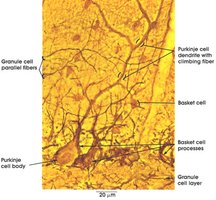
The cerebellum contains Purkinje cells and other specialized neurons.
Summary Table: Types of Neuroglia
Cell Type | Location | Main Function |
|---|---|---|
Astrocytes | CNS | Support, blood-brain barrier, regulate environment |
Microglia | CNS | Defense, phagocytosis |
Ependymal Cells | CNS | Produce/circulate CSF |
Oligodendrocytes | CNS | Myelinate CNS axons |
Schwann Cells | PNS | Myelinate PNS axons |
Satellite Cells | PNS | Support neuron cell bodies |
Key Equations
Resting Membrane Potential
The resting membrane potential is determined by ion concentrations and membrane permeability.
Equation:
Saltatory Conduction
Action potentials jump from node to node, increasing conduction velocity.
Additional info:
Neurons and neuroglia are fundamental to nervous system function and histology.
Myelination is essential for rapid signal transmission.
Connective tissue coverings protect and organize nerves.
Synapses are critical for communication between neurons and other cells.
