 Back
BackLymphatic and Respiratory Systems: Structure and Function
Study Guide - Smart Notes
 Tailored notes based on your materials, expanded with key definitions, examples, and context.
Lymphatic System: Structure and Function
Circulatory vs. Cardiovascular vs. Lymphatic Vessels
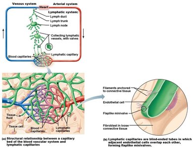
The human body contains several types of vessels that transport fluids. Understanding the differences between these vessels is essential for grasping the organization of the circulatory and lymphatic systems.
Circulatory vessels: A generic term that can refer to both cardiovascular and lymphatic vessels.
Cardiovascular vessels: Only carry blood (arteries, veins, capillaries).
Lymphatic vessels: Carry lymph, a fluid derived from interstitial fluid. Lymphatic capillaries are blind-ended and have overlapping endothelial cells that form mini-valves. Lymphatic vessels generally lack tunics but possess valves to prevent backflow.
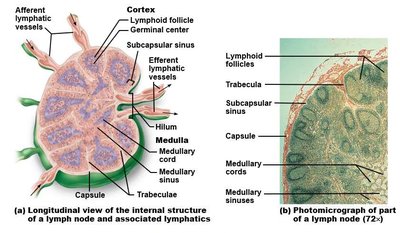
Lymph Nodes vs. Lymph Nodules
Lymph nodes and lymph nodules are both lymphoid structures, but they differ in location, structure, and function.
Lymph nodes: Located along lymphatic vessels, filter lymph before it returns to the venous system. They have a capsule, multiple afferent vessels, and fewer efferent vessels.
Lymph nodules: Do not have a capsule and are found within the submucosal layer of mucous membranes (MALT).
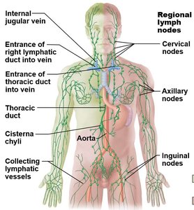
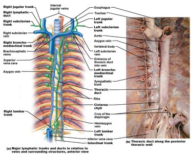
Lymph Drainage and Major Lymphatic Vessels
Lymph is collected from tissues and returned to the bloodstream via a network of lymphatic vessels, trunks, and ducts. Lymph nodes are clustered in specific regions, and lymph is ultimately drained by two main ducts.
Superficial lymph node clusters: Cervical, axillary, and inguinal regions.
Deep lymph node clusters: Tracheobronchial, aortic, and iliac regions.
Lymphatic trunks: Jugular, subclavian, bronchomediastinal, intestinal (unpaired), and lumbar trunks collect lymph from various regions and drain into ducts.
Lymphatic ducts:
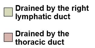
Thoracic duct: Drains the entire left half of the body, right pelvis, and right lower limb.
Right lymphatic duct: Drains the right upper quadrant of the body.
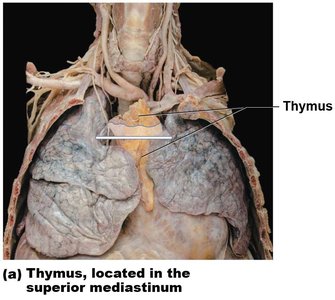
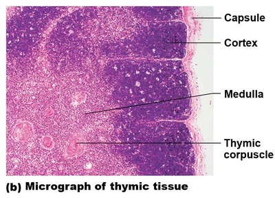
Lymphoid Organs: Thymus
The thymus is a primary lymphoid organ essential for T-cell maturation. T-lymphocytes are produced in the red bone marrow and migrate to the thymus, where they differentiate and mature.
The thymus is divided into lobules, each with a cortex and medulla.
Thymic corpuscles are found in the medulla and are involved in T-cell development.
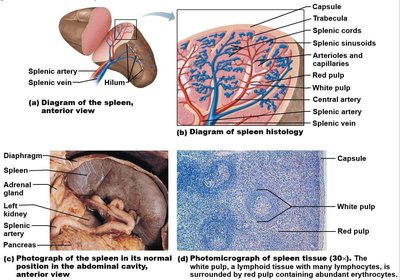
Lymphoid Organs: Spleen
The spleen is the largest lymphoid organ, located in the upper left quadrant (ULQ) of the abdomen, posterior to the stomach. It filters blood, removes old erythrocytes, and mounts immune responses.
Supplied by the splenic artery and vein, which enter at the hilum.
Surrounded by a capsule with inward extensions called trabeculae.
Red pulp: Contains blood-filled sinusoids and cords, responsible for filtering blood.
White pulp: Lymphoid tissue involved in immune responses.
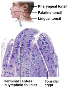
Lymphoid Organs: Tonsils
Tonsils are collections of lymphoid tissue located in the pharynx. They protect against pathogens entering through the oral and nasal cavities.
Tonsils have crypts surrounded by lymphoid follicles.
Types of tonsils:
Palatine tonsils: Lateral sides of the pharynx
Lingual tonsil: Posterior tongue
Pharyngeal tonsil: Roof of pharynx
Tubal tonsils: At the opening of the pharyngotympanic tubes
Respiratory System: Structure and Function
Respiratory Tracts: Upper (Conducting) vs. Lower (Respiratory) Zones
The respiratory system is divided into conducting and respiratory zones. The primary function is ventilation and external respiration.
Conducting zone: Warms, moistens, and transports air; no gas exchange occurs. Includes the nasal cavity, pharynx, larynx, trachea, bronchi, and terminal bronchioles.
Respiratory zone: Site of gas exchange; includes respiratory bronchioles, alveolar ducts, and alveoli.

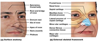
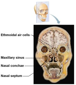
Conducting Zone: Nasal Cavity
The nasal cavity is the entryway for air and is responsible for filtering, warming, and humidifying inhaled air.
Formed by the frontal, nasal, and maxillary bones, and hyaline cartilage plates.
Divided by the nasal septum (ethmoid, vomer, septal cartilage).
The palate separates the nasal and oral cavities (hard and soft palate).
Lined with olfactory and respiratory mucosa.
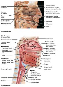

Conducting Zone: Pharynx
The pharynx is a muscular chamber shared by the respiratory and digestive tracts. It is divided into three regions:
Nasopharynx: Air passage only; closed off during swallowing by the uvula and soft palate.
Oropharynx: Extends from the soft palate to the epiglottis.
Laryngopharynx: Posterior to the larynx, leads to the esophagus and larynx.
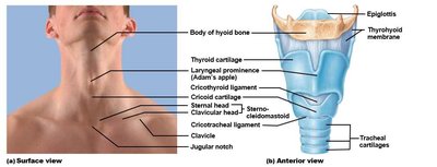
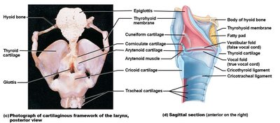
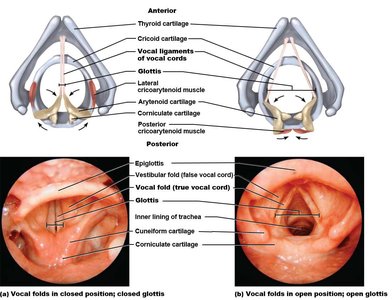
Conducting Zone: Larynx
The larynx is the passageway for air between the pharynx and trachea. It is supported by nine cartilages and is responsible for sound production.
Superiorly attaches to the hyoid bone.
Epiglottis closes the opening to the trachea during swallowing.
Major cartilages: thyroid, cricoid, arytenoid, corniculate, cuneiform, and epiglottis.
Vocal folds (true vocal cords) and vestibular folds (false vocal cords) are involved in sound production and airway protection.
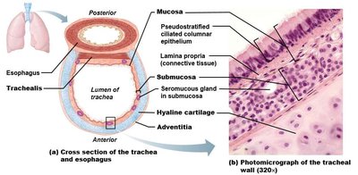
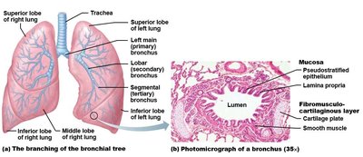
Conducting Zone: Trachea
The trachea is a flexible tube that conducts air to the bronchi. It is supported by C-shaped rings of hyaline cartilage, which keep the airway open while allowing the esophagus to expand during swallowing.
Lined with pseudostratified ciliated columnar epithelium.
Bifurcates at the carina (T7 vertebra) into right and left primary bronchi.
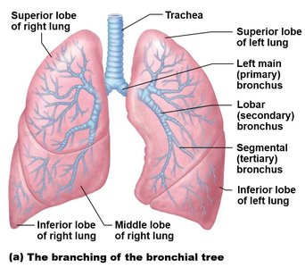
Conducting Zone: Bronchi and Bronchioles
The bronchi branch repeatedly within the lungs, forming the bronchial tree. Cartilage plates diminish as the airways branch, and smooth muscle becomes more prominent. The terminal bronchioles mark the end of the conducting zone.
Primary bronchi supply each lung; secondary (lobar) bronchi supply each lobe; tertiary (segmental) bronchi supply bronchopulmonary segments.
Bronchioles lack cartilage and have abundant smooth muscle.
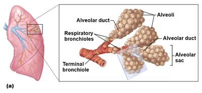
Respiratory Zone: Alveoli and Gas Exchange
The respiratory zone begins with respiratory bronchioles and includes alveolar ducts and alveoli, which are the primary sites of gas exchange.
Type I alveolar cells: Simple squamous cells, form the structure of the alveolar wall.
Type II alveolar cells: Produce surfactant to reduce surface tension and prevent alveolar collapse.
Thoracic Cavity: Gross Anatomy of the Lungs
Each lung is contained within a double-layered pleural membrane. The left lung is smaller due to the cardiac notch and has two lobes, while the right lung has three lobes.
Costal surfaces face the ribs; the apex is deep to the clavicle; the base rests on the diaphragm.
The root of the lung contains bronchi, blood vessels, and nerves.
Lung | Lobes | Fissures |
|---|---|---|
Left | 2 (superior, inferior) | Oblique |
Right | 3 (superior, middle, inferior) | Horizontal, Oblique |
