 Back
BackMuscle Tissue and Physiology: Structure and Function
Study Guide - Smart Notes
 Tailored notes based on your materials, expanded with key definitions, examples, and context.
Introduction to Muscles and Muscle Tissue
Properties of Muscle Tissue
Muscle tissue is specialized for contraction to create movement. It converts chemical energy (from ATP) into mechanical energy, generating force and movement. Muscle tissue also produces heat as a byproduct of contraction.
Contractility: The ability of muscle tissue to forcibly shorten, producing movement or tension.
Extensibility: The ability to be stretched without being damaged.
Elasticity: The ability to return to original length after being stretched or contracted.
Excitability: The ability to respond to stimuli, usually from the nervous system.
Example: Contractility is the property most directly related to the conversion of chemical energy to mechanical energy.
Types of Muscle Tissue
Classification and Characteristics
There are three types of muscle tissue in the human body, each with distinct locations, control mechanisms, and microscopic features.
Muscle Type | Location | Voluntary/Involuntary | Striated | Nuclei per Cell |
|---|---|---|---|---|
Skeletal Muscle | Connected to bones | Voluntary | Striated | Many |
Cardiac Muscle | Heart | Involuntary | Striated | One |
Smooth Muscle | Walls of hollow organs & blood vessels | Involuntary | Non-striated | One |
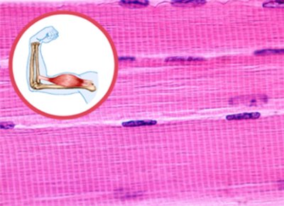
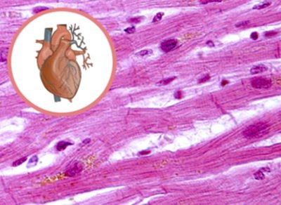
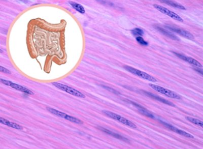
Example: The heart is composed of cardiac muscle, which is involuntary. Therefore, you cannot consciously lower your heart rate in the same way you can flex your biceps.
Practice: If you do not see striations under the microscope, you are likely looking at smooth muscle.
Structure of a Skeletal Muscle
Organization of Muscle Tissue
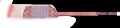
Skeletal muscles are complex organs composed of muscle fibers, nerves, blood vessels, and connective tissue. They are organized into bundles for efficient force transmission and control.
Muscle Fiber: A single, long, multinucleated cell; surrounded by endomysium.
Fascicle: A bundle of muscle fibers; surrounded by perimysium.
Muscle: A bundle of fascicles; surrounded by epimysium.
Connective Tissue Attachments: Tendon (cord-like) and aponeurosis (sheet-like) connect muscle to bone.
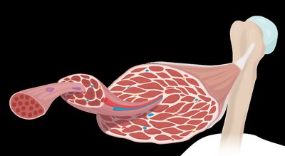
Example: Marbling in beef is due to fat in the connective tissue layers (endomysium, perimysium, epimysium).
Muscle Fiber Structure
Each muscle fiber contains specialized structures for contraction:
Sarcolemma: The plasma membrane of a muscle fiber.
T-Tubules: Invaginations of the sarcolemma that conduct action potentials deep into the fiber.
Myofibrils: Long, rod-shaped organelles containing contractile proteins (actin and myosin).
Sarcoplasmic Reticulum (SR): Specialized endoplasmic reticulum that stores and releases calcium ions (Ca2+).
Sliding Filament Theory and the Sarcomere
Sarcomere Structure
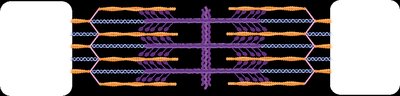
The sarcomere is the smallest contractile unit of muscle, composed of overlapping thick (myosin) and thin (actin) filaments. The arrangement of these filaments creates the striated appearance of skeletal and cardiac muscle.
Myosin: Thick filament, anchored at the M line (center of sarcomere).
Actin: Thin filament, anchored at the Z disc (ends of sarcomere).
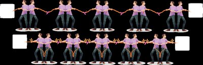
During contraction: Filaments slide past each other, increasing overlap; the sarcomere shortens, but the filaments themselves do not change length.
Proteins of the Sarcomere
Contractile Proteins: Myosin (thick), Actin (thin).
Regulatory Proteins: Tropomyosin (blocks myosin binding sites on actin), Troponin (binds Ca2+ and moves tropomyosin).
Structural Proteins: Titin (provides elasticity and structural support).

Example: If troponin cannot bind calcium, myosin binding sites remain blocked and contraction cannot occur.
Sarcomere Bands, Zones, and Lines
The sarcomere contains distinct regions visible under a microscope:
I Band: Contains only actin (thin filaments).
A Band: Contains both actin and myosin (overlap region).
H Zone: Center of A band with only myosin.
Z Disc: Boundary of the sarcomere; anchors actin.
M Line: Center of the sarcomere; anchors myosin.
Component | Change During Contraction |
|---|---|
A Band | No change |
I Band | Shortens |
H Zone | Shortens |
Z Disc | Move closer together |
M Line | No change |
Steps of Muscle Contraction
Overview of Muscle Contraction
Muscle contraction is initiated by a signal from the nervous system and involves several key steps:
Excitation: A motor neuron stimulates the muscle fiber at the neuromuscular junction, generating an action potential.
Excitation-Contraction Coupling: The action potential travels along the sarcolemma and T-tubules, triggering Ca2+ release from the sarcoplasmic reticulum.
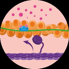
Contraction: Ca2+ binds to troponin, moving tropomyosin and exposing myosin binding sites on actin. Myosin binds actin, forming cross-bridges and producing the power stroke.
Relaxation: Ca2+ is pumped back into the SR, binding sites are covered, and the muscle relaxes.
Neurotransmitters and Action Potentials
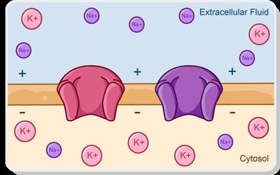
At the neuromuscular junction, the neurotransmitter acetylcholine (ACh) is released, initiating an action potential in the muscle fiber. The action potential is a rapid change in membrane potential due to the movement of Na+ and K+ ions.
Depolarization: Na+ enters the cell, making the inside more positive.
Repolarization: K+ exits the cell, restoring the negative charge inside.
Events at the Neuromuscular Junction
Action potential arrives at the axon terminal.
Voltage-gated Ca2+ channels open; Ca2+ enters the axon terminal.
ACh is released into the synaptic cleft.
ACh binds to receptors on the motor end plate, opening Na+ channels and generating an action potential in the muscle fiber.
ACh is broken down by acetylcholinesterase, ending the signal.
Excitation-Contraction Coupling
This process links the action potential to muscle contraction:
Action potential spreads along the sarcolemma and into T-tubules.
Voltage-gated channels in the sarcoplasmic reticulum release Ca2+ into the sarcomere.
Ca2+ binds to troponin, moving tropomyosin and exposing myosin binding sites on actin.
Myosin binds to actin, forming cross-bridges and initiating contraction.
Cross Bridge Cycle
The cross bridge cycle describes the interaction of actin and myosin during contraction, powered by ATP:
Myosin head binds to actin (cross-bridge formation).
Power stroke: Myosin head pivots, pulling actin and releasing ADP + Pi.
ATP binds to myosin, causing it to detach from actin.
ATP is hydrolyzed, re-cocking the myosin head for another cycle.
Equation:
Example: If a muscle runs out of ATP, myosin cannot detach from actin, resulting in rigor (as seen in rigor mortis).
Additional info: The notes above expand on the original content by providing definitions, examples, and logical academic context for clarity and completeness.
