 Back
BackNervous System: Development, Anatomy, and Function
Study Guide - Smart Notes
 Tailored notes based on your materials, expanded with key definitions, examples, and context.
Nervous System Development, Anatomy, and Function
Embryonic Development of the Nervous System
The nervous system (NS) originates from the embryonic ectoderm, the outermost germ layer that also forms the skin. Early in development, the central region of the embryo's back forms the neural plate with raised neural folds. These folds converge and fuse, creating the neural tube (precursor to the CNS) and the neural crest (precursor to the PNS and other structures).
Neural Crest: Gives rise to peripheral neuron cells, adrenal medulla, melanocytes, meninges, and PNS glial cells.
Neural Tube: Forms CNS neuron cells and CNS glial cells.
Example: Failure of neural tube closure can result in neural tube defects such as spina bifida.
Basic Design of the Central Nervous System (CNS)
The CNS is organized into distinct layers and regions, each with specialized functions:
Central Lumen: Filled with cerebrospinal fluid (CSF).
Gray Matter: Contains neuron cell bodies; forms the cortex in the cerebrum and cerebellum, and nuclei within the brain and spinal cord.
White Matter: Composed of myelinated axons; responsible for communication between different CNS regions.
Meninges: Protective coverings of the CNS (dura mater, arachnoid mater, pia mater).
CSF Formation and Flow
Cerebrospinal fluid (CSF) is produced by the choroid plexus in the brain's ventricles. It circulates through the ventricular system and into the subarachnoid space, providing mechanical and chemical protection for the brain and spinal cord.
Functions: Cushions the brain, maintains ionic balance, provides limited nutrition, and removes waste.
Flow: CSF flows from the ventricles into the subarachnoid space and is reabsorbed into the venous system via arachnoid villi.
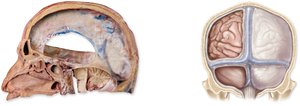
Meninges
The meninges are three connective tissue membranes that protect the brain and spinal cord:
Dura Mater: Outermost, tough layer; forms venous sinuses and partitions (falx cerebri, tentorium cerebelli).
Arachnoid Mater: Middle, web-like layer; contains the subarachnoid space filled with CSF.
Pia Mater: Innermost, delicate layer; adheres closely to the brain and spinal cord.
Example: Inflammation of the meninges is called meningitis.
Arterial Blood Supply and Venous Drainage of the Brain
The brain receives blood from the internal carotid and vertebral arteries, which form the Circle of Willis—an arterial anastomosis ensuring continuous blood flow. Venous blood drains through dural venous sinuses into the internal jugular veins.
Choroid Plexus: Site of CSF production; also involved in nutrient and waste exchange.
Circumventricular Organs: Specialized brain regions lacking a blood-brain barrier, involved in homeostatic regulation.
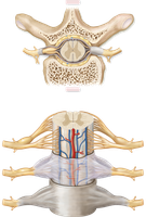
Anatomy of the Spinal Cord
The spinal cord is organized into gray and white matter, with enlargements for limb innervation and specialized structures at its inferior end.
Gray Matter: Contains neuron cell bodies; organized into anterior (motor), posterior (sensory), and lateral (autonomic) horns.
White Matter: Contains ascending (sensory) and descending (motor) tracts.
Special Structures: Cervical and lumbar enlargements, conus medullaris, cauda equina, filum terminale.
Functions of the Spinal Cord
The spinal cord conducts information between the body and brain and mediates reflexes.
Conduction: Sensory and motor pathways.
Reflexes: Rapid, involuntary responses (e.g., withdrawal reflex, cross extensor reflex).
Brainstem: Medulla Oblongata, Pons, and Midbrain
The brainstem connects the spinal cord to higher brain centers and controls vital functions.
Medulla Oblongata: Controls cardiovascular, respiratory, and digestive functions; site of pyramidal decussation.
Pons: Relays information, regulates breathing, connects cerebellar hemispheres.
Midbrain: Contains nuclei for visual and auditory reflexes, and motor coordination.
Cerebellum
The cerebellum coordinates voluntary movements, balance, and posture. It receives sensory input about body position and motor commands, integrating them to fine-tune movements.
Lobes: Anterior and posterior.
Connections: Receives input from the spinal cord, cerebral cortex, vestibular system, and other brain regions.
Diencephalon: Thalamus, Hypothalamus, Epithalamus
The diencephalon is a central brain region involved in sensory relay, homeostasis, and endocrine regulation.
Thalamus: Major sensory relay station; involved in motor and emotional processing.
Hypothalamus: Regulates the autonomic nervous system, endocrine system, emotions, hunger, thirst, temperature, and circadian rhythms.
Epithalamus: Contains the pineal gland, which secretes melatonin for circadian rhythm regulation.
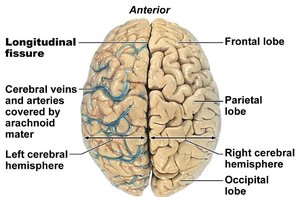
Cerebrum
The cerebrum is the largest part of the brain, responsible for higher cognitive functions, voluntary movement, and sensory perception. It is divided into lobes and hemispheres, each with specialized functions.
Lobes: Frontal (motor, executive), parietal (sensory), temporal (auditory, memory), occipital (visual).
Tracts: Association (within hemisphere), commissural (between hemispheres), projection (to/from lower CNS).
Gyri: Pre-central (motor), post-central (sensory).
Specialized Areas of the Cerebrum
Motor Areas: Pre-central gyrus, premotor cortex, Broca’s area (speech).
Sensory Areas: Post-central gyrus, visual cortex (occipital), auditory cortex (temporal), Wernicke’s area (language comprehension).
Basal Ganglia (Nuclei): Unconscious integration of muscle movements.
Limbic System: Emotional processing, memory (hippocampus), and motivation.
Diseases and Injuries of the Nervous System
Several disorders can affect the nervous system, impacting function and health:
Hydrocephalus: Accumulation of CSF due to impaired drainage.
Seizures: Abnormal, excessive neuronal activity.
Concussion: Temporary loss of brain function due to trauma.
TIA/CVA (Stroke): Loss of blood flow to brain tissue, causing neuron death.
Dementia: Progressive loss of memory and cognitive function (e.g., Alzheimer’s disease).
Parkinson’s Disease: Motor disorder due to basal nuclei dysfunction and dopamine deficiency.
