Back
BackNervous Tissue: Structure, Function, and Physiology
Study Guide - Smart Notes
 Tailored notes based on your materials, expanded with key definitions, examples, and context.
Nervous Tissue and the Nervous System
Overview and Functions
The nervous system is responsible for controlling and integrating all body activities within limits that maintain life. It performs three basic functions:
Sensory Function: Detects changes in the internal and external environment using sensory receptors.
Integrative Function: Interprets and remembers sensory input, making decisions about appropriate responses.
Motor Function: Responds to stimuli by activating effectors such as muscles and glands.
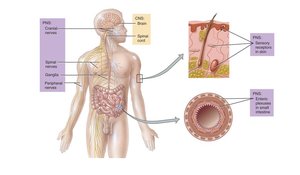
The major structures of the nervous system include the brain, cranial nerves, spinal cord, spinal nerves, ganglia, enteric plexuses, and sensory receptors.
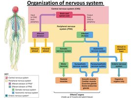
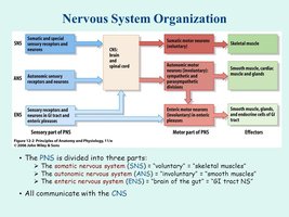
Organization of the Nervous System
The nervous system is divided into the central nervous system (CNS) and the peripheral nervous system (PNS). The CNS consists of the brain and spinal cord, while the PNS includes cranial and spinal nerves, ganglia, and sensory receptors. The PNS is further subdivided into the somatic, autonomic, and enteric nervous systems.
Neurons and Neuroglia
Neurons: Structure and Function
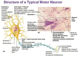
Neurons are the functional units of the nervous system. They are specialized for the reception, processing, and transmission of information through electrical impulses called action potentials. Most neurons do not divide, making them a limited resource in the body.
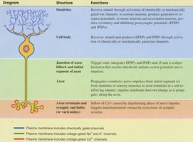
Dendrites: Conduct impulses toward the cell body; typically short, branched, and unmyelinated.
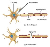
Cell Body (Soma): Contains the nucleus, Nissl bodies, and other organelles.
Axon: Conducts impulses away from the cell body; arises from the axon hillock and ends in synaptic terminals containing neurotransmitters.
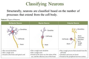
Classification of Neurons
Neurons can be classified based on structure and function:
Multipolar Neurons: Several dendrites and one axon; most common in the CNS.
Bipolar Neurons: One main dendrite and one axon; found in the retina, inner ear, and olfactory area.
Unipolar Neurons: Single process that divides into two branches; found in sensory neurons of the PNS.
Type | Structure | Location |
|---|---|---|
Multipolar | Several dendrites, one axon | Brain, spinal cord, motor neurons |
Bipolar | One dendrite, one axon | Retina, inner ear, olfactory area |
Unipolar | Single process divides into two branches | Sensory neurons in PNS |
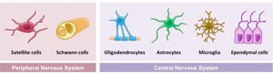
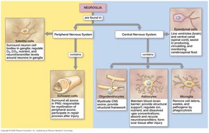
Neuroglia (Glial Cells)
Neuroglia are supportive cells that do not conduct action potentials. They maintain the environment for neurons, provide structural support, and participate in repair processes. Neuroglia are more numerous than neurons and can divide, which is significant in the formation of tumors (gliomas).
Astrocytes: Maintain the blood-brain barrier and provide structural support.
Oligodendrocytes: Myelinate axons in the CNS.
Microglia: Act as phagocytes in the CNS.
Ependymal Cells: Line ventricles and produce cerebrospinal fluid.
Schwann Cells: Myelinate axons in the PNS.
Satellite Cells: Support neuron cell bodies in the PNS.
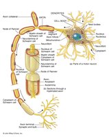
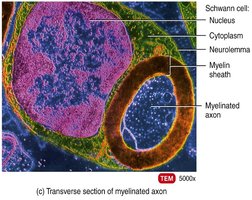
Myelination and Axon Coverings
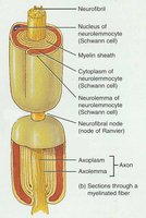
Myelin Sheath
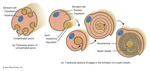
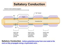
The myelin sheath is a lipid and protein covering that insulates axons and increases the speed of nerve impulse conduction. In the PNS, Schwann cells form the myelin sheath, while in the CNS, oligodendrocytes perform this function. Gaps in the myelin sheath, called nodes of Ranvier, are crucial for rapid signal transmission.
Myelinated Fibers: Fast conduction, appear white.
Unmyelinated Fibers: Slow conduction, only surrounded by neurilemma.
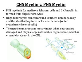
Myelination in CNS vs. PNS
Oligodendrocytes myelinate axons in the CNS, but unlike Schwann cells, their cell bodies do not surround the axons, and no neurilemma is formed. This limits the ability of CNS axons to regenerate after injury. In contrast, Schwann cells in the PNS can guide axonal regrowth.
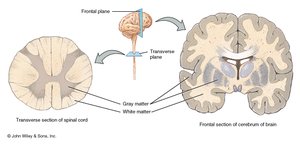
Gray and White Matter
White matter consists of myelinated axons, while gray matter contains neuron cell bodies, dendrites, unmyelinated axons, and neuroglia. In the spinal cord, gray matter forms an H-shaped core, while in the brain, it forms a thin outer shell and clusters called nuclei.
Electrical Signals in Neurons
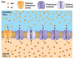
Resting Membrane Potential (RMP)
Neurons are excitable due to the voltage difference across their membranes, primarily established by the unequal distribution of sodium (Na+) and potassium (K+) ions. The RMP is typically -70 mV, with the inside of the cell being more negative than the outside.
Na+/K+ Pump: Maintains the RMP by pumping 3 Na+ out and 2 K+ in.
Leakage Channels: More permeable to K+ than Na+, contributing to the negative RMP.
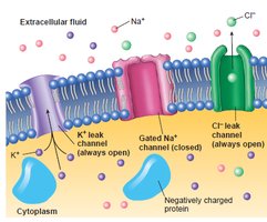
Types of Ion Channels
Leakage (Non-gated) Channels: Always open; more K+ than Na+ channels.
Gated Channels: Open in response to stimuli (voltage, chemicals, mechanical, or light).
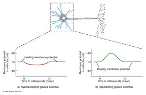
Graded Potentials
Graded potentials are small, localized changes in membrane potential caused by the opening of gated ion channels. They can be depolarizing (more positive) or hyperpolarizing (more negative) and vary in amplitude depending on the strength of the stimulus.
Action Potentials
An action potential is a rapid, all-or-none electrical event that propagates along the axon. It is initiated when graded potentials reach a threshold, typically -55 mV. The process involves:
Depolarization: Voltage-gated Na+ channels open, Na+ enters the cell, making the inside more positive.
Repolarization: Na+ channels close, K+ channels open, K+ exits the cell, restoring negativity.
After-hyperpolarization: Membrane potential may become more negative than RMP before returning to baseline.


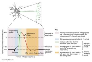
Refractory Period
The refractory period is the time during which a neuron cannot generate another action potential. It includes:
Absolute Refractory Period: No stimulus can initiate another action potential.
Relative Refractory Period: A stronger-than-normal stimulus can initiate another action potential.

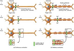
Conduction of Action Potentials
Action potentials propagate along axons by two mechanisms:
Continuous Conduction: Occurs in unmyelinated fibers; step-by-step depolarization along the entire axon.
Saltatory Conduction: Occurs in myelinated fibers; action potentials jump from node to node, increasing speed.
Synaptic Transmission
Chemical Synapses
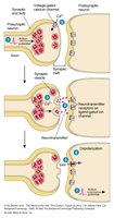
At chemical synapses, the action potential triggers the opening of voltage-gated Ca2+ channels, leading to neurotransmitter release. The neurotransmitter crosses the synaptic cleft and binds to receptors on the postsynaptic cell, causing a change in membrane potential.

Removal of Neurotransmitter
Neurotransmitters are removed from the synaptic cleft by:
Diffusion: Moving away from the synapse into the bloodstream.
Enzymatic Degradation: Breakdown by enzymes (e.g., acetylcholinesterase).
Uptake: Reuptake by neurons or glial cells.
Neurotransmitter Effects
Agonists: Enhance neurotransmitter effects (e.g., drugs that mimic neurotransmitters).
Antagonists: Block neurotransmitter action (e.g., drugs that block receptors or release).
Repair and Disorders of Nervous Tissue
Repair in the PNS
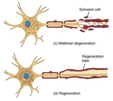
Axons and dendrites in the PNS can regenerate if the cell body is intact and Schwann cells form a regeneration tube. The process involves chromatolysis, Wallerian degeneration, and axonal regrowth.
Local Anesthetics
Local anesthetics such as novocaine and lidocaine block sodium channels, preventing action potential generation and thus blocking pain sensation.

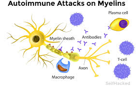
Multiple Sclerosis (MS)
MS is an autoimmune disorder that destroys myelin sheaths in the CNS, leading to scar formation and progressive loss of function. Symptoms include muscle weakness, abnormal sensations, and vision problems.
Epilepsy
Epilepsy is characterized by recurrent seizures due to abnormal electrical discharges in the brain. Causes include brain injury, metabolic disturbances, infections, toxins, and tumors.
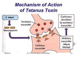
Axonal Transport and Disease
Toxins and pathogens can use axonal transport to reach neuron cell bodies. Examples include tetanus toxin (causing muscle spasms) and viruses such as rabies and herpes, which can travel to the CNS.