 Back
BackThe Cellular Level of Organization: Structure and Function of Cells
Study Guide - Smart Notes
 Tailored notes based on your materials, expanded with key definitions, examples, and context.
The Cellular Level of Organization
An Introduction to Cells
Cells are the fundamental units of life, responsible for carrying out all essential physiological functions. The study of cell structure and function is known as cytology. According to cell theory, all living organisms are composed of cells, all cells arise from preexisting cells, and cells are the smallest units capable of performing life's essential functions. In the human body, there are two major cell types: sex cells (sperm and oocytes) and somatic cells (all other body cells).
The Plasma Membrane
Structure and Function
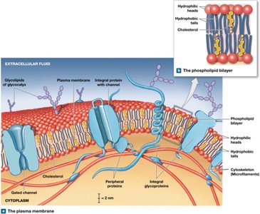
The plasma membrane forms the outer boundary of the cell, providing selective transport of substances and maintaining cellular integrity. It is primarily composed of a phospholipid bilayer with embedded proteins and carbohydrates. The membrane's structure allows it to perform several critical functions:
Physical isolation: Separates the cytoplasm from the extracellular environment.
Regulation of exchange: Controls the entry and exit of ions, nutrients, and wastes.
Sensitivity: Contains receptors that detect chemical signals.
Structural support: Anchors cells to each other and to extracellular materials.
Membrane Components
Phospholipids: Form a bilayer with hydrophilic heads facing outward and hydrophobic tails inward, creating a barrier to water-soluble substances.
Cholesterol: Stabilizes membrane fluidity and permeability.
Proteins: Integral (span the membrane) and peripheral (bound to surfaces); function as anchors, receptors, enzymes, carriers, and channels.
Carbohydrates: Glycoproteins and glycolipids form the glycocalyx, involved in cell recognition, protection, and adhesion.
Organelles
Cytoplasm and Nonmembranous Organelles
The cytoplasm includes all material between the plasma membrane and the nuclear envelope. It consists of cytosol (fluid) and organelles (specialized structures). Nonmembranous organelles are not surrounded by membranes and include:
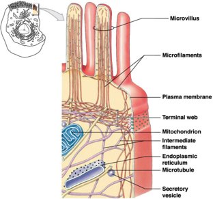
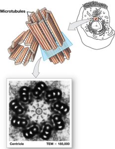
Cytoskeleton: Provides structural support and facilitates movement. Composed of microfilaments (actin), intermediate filaments, and microtubules (tubulin).
Microvilli: Finger-like projections that increase surface area for absorption.
Centrioles: Organize microtubules during cell division.
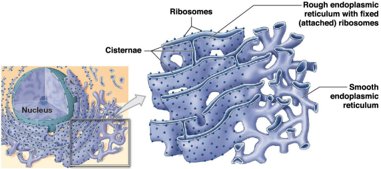
Ribosomes: Sites of protein synthesis; can be free or attached to the rough ER.
Proteasomes: Degrade damaged or unneeded proteins.
Membranous Organelles
Endoplasmic Reticulum (ER): Network of membranes involved in synthesis, storage, and transport. Rough ER (with ribosomes) synthesizes proteins; Smooth ER (without ribosomes) synthesizes lipids and detoxifies chemicals.
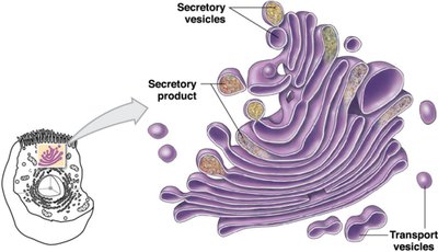
Golgi Apparatus: Modifies, sorts, and packages proteins and lipids for secretion or delivery to other organelles.
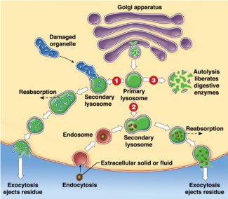
Lysosomes: Vesicles containing digestive enzymes; break down waste, damaged organelles, and foreign materials. Involved in autolysis (self-destruction of damaged cells).
Peroxisomes: Vesicles containing enzymes that break down fatty acids and neutralize toxic compounds (e.g., hydrogen peroxide).
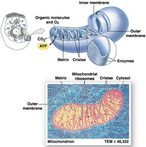
Mitochondria: Double-membraned organelles that produce ATP through aerobic respiration. Contain their own DNA and ribosomes.
The Nucleus
Structure and Function
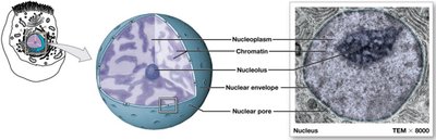
The nucleus is the control center of the cell, containing genetic material (DNA) and directing cellular activities. It is surrounded by a double membrane called the nuclear envelope, which contains nuclear pores for molecular exchange. The nucleoplasm contains chromatin (DNA and proteins), the nucleolus (site of rRNA synthesis), and the nuclear matrix (structural support).
Genetic Code and Protein Synthesis
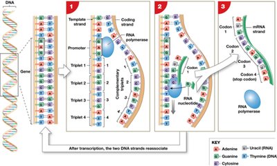
The genetic code is the sequence of nucleotide bases (A, T, C, G) in DNA that encodes instructions for protein synthesis. Genes are segments of DNA that code for specific proteins. Protein synthesis involves two main processes:
Transcription: DNA is used as a template to synthesize messenger RNA (mRNA) in the nucleus.
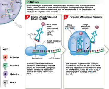
Translation: mRNA is decoded by ribosomes in the cytoplasm to assemble amino acids into a polypeptide chain. Transfer RNA (tRNA) brings amino acids to the ribosome, matching codons on mRNA with anticodons on tRNA.
Membrane Transport Mechanisms
Diffusion and Osmosis
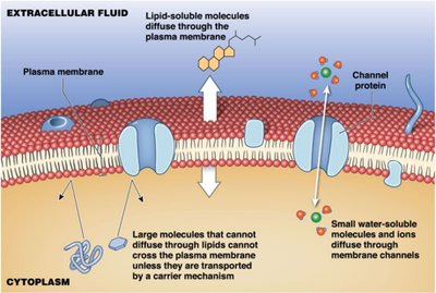
The plasma membrane is selectively permeable, allowing certain substances to cross while restricting others. Transport mechanisms include:
Diffusion: Movement of molecules from high to low concentration. Includes simple diffusion (lipid-soluble molecules) and channel-mediated diffusion (water-soluble molecules and ions).
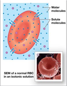
Osmosis: Diffusion of water across a selectively permeable membrane toward higher solute concentration. Osmotic pressure drives water movement; tonicity describes the effect of solutions on cell volume (isotonic, hypotonic, hypertonic).
Carrier-Mediated and Active Transport
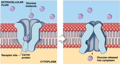
Facilitated diffusion: Passive transport of large or non-lipid-soluble molecules via carrier proteins.
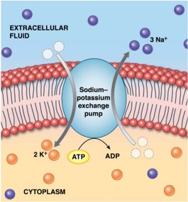
Active transport: Movement of substances against their concentration gradient using energy (ATP). The sodium-potassium pump exchanges 3 Na+ out for 2 K+ in per ATP hydrolyzed.
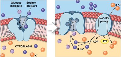
Secondary active transport: Uses the gradient established by primary active transport (e.g., Na+ gradient) to drive the transport of other molecules (e.g., glucose cotransport).
Vesicular Transport
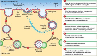
Vesicular (bulk) transport moves large particles or volumes via vesicles and requires ATP. Types include:
Endocytosis: Import of materials into the cell (receptor-mediated, pinocytosis, phagocytosis).
Exocytosis: Export of materials out of the cell.
Cell Membrane Potential
Origin and Significance
The membrane potential is the voltage difference across the plasma membrane, primarily due to the unequal distribution of Na+ and K+ ions. The resting membrane potential (about -70 mV) is maintained by the sodium-potassium pump and leak channels. This potential is essential for electrical signaling in neurons and muscle cells and for secondary active transport.
The Cell Life Cycle
Stages and Regulation
The cell life cycle includes interphase (G1, S, G2 phases) and the mitotic phase (mitosis and cytokinesis). The cycle is regulated by checkpoints (G1, G2, M) and proteins such as cyclins and cyclin-dependent kinases (Cdks). Uncontrolled cell division can lead to cancer, involving mutations in proto-oncogenes and tumor suppressor genes (e.g., p53, Rb).
Cellular Differentiation
Definition and Importance
Cellular differentiation is the process by which cells become specialized in structure and function. Although all cells contain the same DNA, different genes are expressed in different cell types, allowing the formation of diverse tissues and organs necessary for multicellular life.
