 Back
BackThe Central Nervous System: Structure, Function, and Integration
Study Guide - Smart Notes
 Tailored notes based on your materials, expanded with key definitions, examples, and context.
Overview of Central Nervous System (CNS) Functions
Functions of the Nervous System
The nervous system is responsible for detecting, integrating, and responding to stimuli both inside and outside the body. It is divided into sensory, integrative, and motor functions:
Sensory Functions: Detection of sensations (performed by the Peripheral Nervous System, PNS)
Integrative Functions: Decision-making processes (performed exclusively by the CNS)
Motor Functions: Stimulation of muscle contractions or gland secretions (performed by the PNS)
Basic Structure of the Brain and Spinal Cord
Brain Anatomy and Divisions
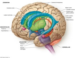
The brain is a soft, whitish-gray organ located in the cranial cavity. It consists of four main divisions and is composed primarily of nervous tissue, with some connective and modified epithelial tissue. Internal cavities called ventricles are filled with cerebrospinal fluid (CSF), which protects and nourishes the brain. The brain receives about 20% of the body's blood flow at rest.
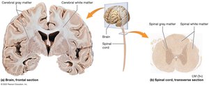
White Matter vs. Gray Matter
White matter contains myelinated axons, while gray matter consists of neuron cell bodies, dendrites, and unmyelinated axons. In the brain, gray matter forms the outer cortex and is scattered in deeper regions as nuclei. In the spinal cord, gray matter is internal, surrounded by superficial white matter tracts.
The Cerebrum
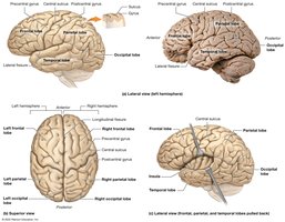
Surface Features and Lobes
The cerebrum is divided into paired hemispheres, separated by the longitudinal fissure. The surface is marked by gyri (ridges), sulci (shallow grooves), and fissures (deep grooves). The main lobes are:
Frontal Lobe: Anterior, bounded posteriorly by the central sulcus
Parietal Lobe: Posterior to the frontal lobe
Temporal Lobe: Lateral surface, separated by the lateral fissure
Occipital Lobe: Posterior, separated by the parieto-occipital sulcus
Insula: Deep to the other lobes, visible when they are separated
Gray Matter: The Cerebral Cortex
The neocortex is the largest part of the cerebral cortex and is responsible for conscious processes. It contains:
Primary Motor Cortex: Plans and executes movement
Primary Sensory Cortices: Process sensory input
Association Areas: Integrate multiple types of stimuli
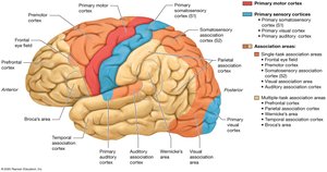
Motor and Sensory Cortices
Motor Cortices: Upper motor neurons in the precentral gyrus (frontal lobe) plan voluntary movement; lower motor neurons in the PNS execute movement.
Somatosensory Areas: Located in the parietal lobe; process touch, temperature, vibration, pressure, stretch, and joint position.
Visual Areas: Occipital lobe; process visual input.
Auditory Areas: Superior temporal lobe; process auditory stimuli.
Other Sensory Areas: Gustatory cortex (taste), vestibular areas (equilibrium), olfactory cortex (smell).
Association Areas and Higher Functions
Language Areas: Broca’s area (speech production, frontal lobe), Wernicke’s area (language comprehension, temporal/parietal lobes)
Prefrontal Cortex: Personality, learning, memory, psychological state
Parietal and Temporal Association Cortices: Integrate sensory stimuli, language, recognition, spatial awareness, and attention
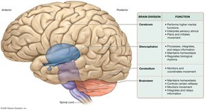
Major Brain Structures and Their Functions
The brain is divided into several major regions, each with specialized functions:
Brain Division | Function |
|---|---|
Cerebrum | Performs higher mental functions, interprets sensory stimuli, plans and initiates movement |
Diencephalon | Processes, integrates, and relays information; maintains homeostasis; regulates biological rhythms |
Cerebellum | Monitors and coordinates movement |
Brainstem | Maintains homeostasis, controls certain reflexes, monitors movement, integrates and relays information |
Sleep and Wakefulness
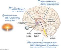
Sleep and Circadian Rhythms
Sleep is a reversible suspension of consciousness that allows the brain to replenish energy and clear metabolic waste. Circadian rhythms regulate the sleep-wake cycle, typically alternating periods of wakefulness and sleep over a 24-hour period.
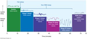
Brain Waves and Stages of Sleep
Brain activity during sleep is measured by an electroencephalogram (EEG), which records brain waves. Sleep progresses through several stages:
Beta Waves: Awake, alert state; low amplitude, high frequency
Theta Waves: Light sleep; amplitude increases, frequency decreases
Delta Waves: Deep sleep; low frequency, high amplitude
REM Sleep: Rapid eye movement, vivid dreams, muscle paralysis, brain waves resemble beta waves
Cognition, Language, Learning, and Memory
Cognition and Cerebral Lateralization
Cognition involves complex tasks such as awareness, reasoning, and personality, primarily managed by the prefrontal cortex. Cerebral lateralization refers to the unequal distribution of cognitive functions between the right and left hemispheres (e.g., language is typically left-lateralized).
Language
Broca’s Area: Production of language, grammar, and syntax (frontal lobe)
Wernicke’s Area: Understanding language and symbolic meaning (temporal lobe)
Aphasia: Language deficit due to damage in these areas
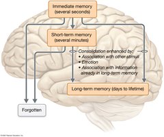
Learning and Memory
Declarative Memory: Facts, readily available to consciousness
Nondeclarative Memory: Skills and associations, largely unconscious
Immediate Memory: Lasts seconds
Short-Term Memory: Lasts minutes
Long-Term Memory: Lasts days to a lifetime; consolidation is the process of transferring information to long-term storage
Protection of the Brain
Cranial Meninges
The brain is protected by three connective tissue membranes called meninges:
Dura Mater: Outermost, toughest layer
Arachnoid Mater: Middle layer; subarachnoid space contains CSF
Pia Mater: Innermost, delicate layer
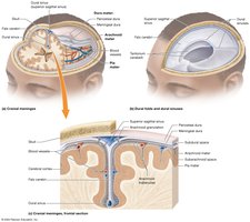
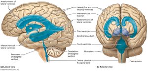
Ventricles and Cerebrospinal Fluid (CSF)
The brain contains four ventricles filled with CSF, which cushions, nourishes, and supports the brain. CSF is produced by the choroid plexuses and circulates through the ventricles and subarachnoid space.
Blood Brain Barrier
The blood brain barrier is formed by endothelial cells with tight junctions, basal laminae, and astrocyte foot processes. It restricts the passage of substances from the blood into the brain, protecting neural tissue from toxins and pathogens.
The Spinal Cord
Structure and Function
The spinal cord serves as a relay station between the brain and the body and as a processing center for spinal reflexes. It is protected by spinal meninges and is anchored within the vertebral cavity.
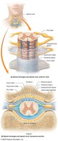
External Anatomy
The spinal cord extends from the foramen magnum to the first or second lumbar vertebra. It features cervical and lumbar enlargements, spinal nerves, and the cauda equina. The conus medullaris marks its terminal end.
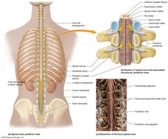
Internal Anatomy
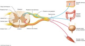
Internally, the spinal cord contains butterfly-shaped gray matter (anterior, posterior, and lateral horns) surrounded by white matter tracts (funiculi). The central canal runs through the center, filled with CSF.
Anterior Horn: Somatic motor functions
Posterior Horn: Somatic and visceral sensory processing
Lateral Horn: Visceral motor control (only in thoracic and lumbar regions)
White Matter Tracts
White matter is organized into posterior, lateral, and anterior funiculi, each containing ascending (sensory) and descending (motor) tracts. These tracts are bilaterally symmetrical and serve both sides of the body.
Sensation and Special Senses
Role of the CNS in Sensation
Sensory stimuli are detected by the PNS and relayed to the CNS, where they are interpreted in the cerebral cortex as conscious perception. Sensations are categorized as general or special senses.
Special Senses
Vision: Processed in the occipital lobe
Hearing: Processed in the temporal lobe
Taste: Processed in the insula and parietal lobes
Smell: Processed in the limbic system and other brain regions
Balance: Involves the brainstem, cerebellum, and thalamus
Movement and Motor Control
Role of the CNS in Voluntary Movement
Voluntary movement is planned and coordinated by the motor areas of the cerebral cortex, basal nuclei, cerebellum, and spinal cord. Three types of neurons are involved:
Upper Motor Neurons: Cell bodies in the cortex or brainstem
Interneurons: In the brainstem and spinal cord
Lower Motor Neurons: Cell bodies in the anterior horn of the spinal cord; axons innervate skeletal muscles
