Back
BackThe Endocrine System: Pituitary, Thyroid, Parathyroid, and Adrenal Glands
Study Guide - Smart Notes
 Tailored notes based on your materials, expanded with key definitions, examples, and context.
The Endocrine System
Overview of Endocrine Glands
The endocrine system is composed of glands that secrete hormones directly into the bloodstream, regulating various physiological processes. Major glands include the pituitary, thyroid, parathyroid, adrenal glands, pancreas, gonads, and pineal gland. Each gland produces specific hormones that target organs and tissues throughout the body.

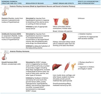
Anterior Pituitary Hormones
Gonadotropins: FSH and LH
Follicle-stimulating hormone (FSH) and Luteinizing hormone (LH) are essential for reproductive function. They are absent in prepubertal children and are regulated by gonadotropin-releasing hormone (GnRH) from the hypothalamus. Gonadal hormones provide negative feedback to suppress their release.
FSH: Stimulates gamete (egg or sperm) production.
LH: Promotes production of gonadal hormones (estrogen, progesterone, testosterone).
Regulation: GnRH triggers release; gonadal hormones inhibit via feedback.
Prolactin (PRL)
Prolactin is secreted by the anterior pituitary and primarily stimulates milk production in females. Its release is inhibited by prolactin-inhibiting hormone (PIH), which is dopamine. Estrogen increases PRL levels, explaining breast changes during the menstrual cycle. Suckling stimulates further PRL release for continued lactation.
Role in males: Not well understood.
Regulation: PIH prevents release until needed; decreased PIH leads to lactation.
Summary Table: Pituitary Hormones
Hormone | Regulation of Release | Target Organ and Effects | Effects of Hypo/Hypersecretion |
|---|---|---|---|
Growth hormone (GH) | Stimulated by GHRH; inhibited by GHIH | Liver, muscle, bone, cartilage; stimulates growth | ↓: Pituitary dwarfism; ↑: Gigantism/acromegaly |
Thyroid-stimulating hormone (TSH) | Stimulated by TRH; inhibited by feedback | Thyroid gland; stimulates thyroid hormone release | ↓: Cretinism/myxedema; ↑: Hyperthyroidism |
Adrenocorticotropic hormone (ACTH) | Stimulated by CRH; inhibited by feedback | Adrenal cortex; stimulates corticosteroid release | ↑: Cushing's disease |
FSH/LH | Stimulated by GnRH; inhibited by feedback | Ovaries/testes; gamete and hormone production | ↓: Failure of sexual maturation |
Prolactin (PRL) | Stimulated by decreased PIH | Breast tissue; promotes lactation | ↓: Poor milk production; ↑: Inappropriate milk production |
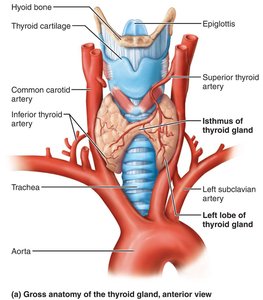
The Thyroid Gland
Anatomy and Histology
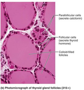
The thyroid gland is a butterfly-shaped organ located in the anterior neck, overlying the trachea. It consists of two lateral lobes connected by an isthmus. The gland contains follicles filled with colloid, surrounded by follicular cells (which secrete thyroid hormone), and parafollicular cells (which secrete calcitonin).
Thyroid Hormone (TH)
Thyroid hormone is the body's major metabolic hormone, existing in two forms: T4 (thyroxine) and T3 (triiodothyronine). T4 is the predominant form in circulation, but T3 is more active and is produced from T4 in peripheral tissues. TH enters target cells, binds to nuclear receptors, and regulates gene transcription, affecting nearly every cell in the body.
Functions: Increases basal metabolic rate (BMR), heat production (calorigenic effect), regulates tissue growth and development, and maintains blood pressure.
BMR Calculation:
Regulation of Thyroid Hormone Release
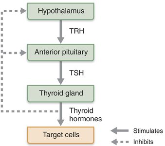
TH release is regulated by a negative feedback loop involving the hypothalamus, anterior pituitary, and thyroid gland. Low TH levels stimulate the release of thyrotropin-releasing hormone (TRH) from the hypothalamus, which triggers thyroid-stimulating hormone (TSH) release from the anterior pituitary, stimulating the thyroid gland to produce TH.
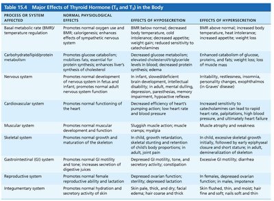
Major Effects of Thyroid Hormone
System | Normal Effects | Hyposecretion | Hypersecretion |
|---|---|---|---|
BMR/Temperature | Increases BMR, heat production | ↓ BMR, cold intolerance, weight gain | ↑ BMR, heat intolerance, weight loss |
Metabolism | Stimulates glucose, fat, protein metabolism | ↓ Metabolism, high cholesterol | ↑ Catabolism, muscle wasting |
Nervous System | Normal development/function | Mental sluggishness, developmental delay | Restlessness, insomnia |
Cardiovascular | Increases heart rate, BP | ↓ HR, ↓ BP | ↑ HR, ↑ BP |
Muscular/Skeletal | Normal growth/function | Weakness, stunted growth | Muscle atrophy, osteoporosis |
GI/Reproductive/Integumentary | Normal motility, fertility, skin/hair health | Constipation, infertility, dry skin/hair | Diarrhea, menstrual irregularity, thin skin/hair |
Thyroid Disorders
Hyposecretion: Myxedema and Goiter
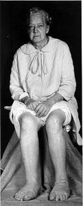
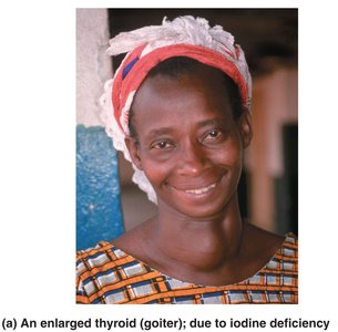
Myxedema is severe hypothyroidism in adults, characterized by low metabolic rate, thick/dry skin, puffy eyes, and lethargy. If caused by iodine deficiency, the thyroid enlarges, forming a goiter. Treatment involves hormone replacement.
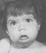
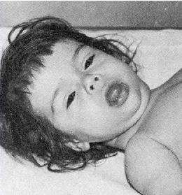
Congenital Hypothyroidism (Cretinism)
Usually due to poor thyroid development, pituitary problems, or maternal factors. Symptoms include weak cry, poor feeding, constipation, and prolonged jaundice. Lifelong TH replacement is necessary.
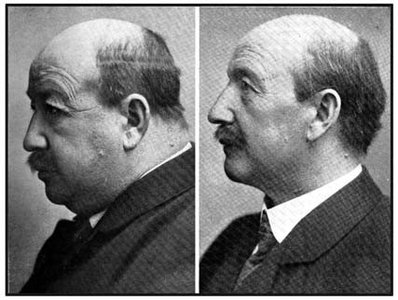
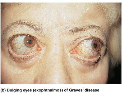
Hypersecretion: Graves' Disease
Graves' disease is an autoimmune disorder where antibodies mimic TSH, causing excessive TH release. Symptoms include high metabolic rate, sweating, rapid heartbeat, nervousness, weight loss, and exophthalmos (bulging eyes). Treatment includes thyroid removal or radioactive iodine.
Calcitonin
Calcitonin is produced by parafollicular (C) cells in response to high blood calcium levels. It inhibits osteoclast activity, reducing calcium release from bone, and stimulates calcium uptake into bone. Its physiological role in humans is minor, but pharmacologically, it is used to treat osteoporosis.

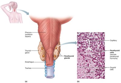
Parathyroid Glands
Anatomy and Function
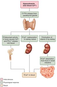
The parathyroid glands are small glands located on the posterior aspect of the thyroid. They secrete parathyroid hormone (PTH), the most important hormone in calcium homeostasis. PTH is released in response to low blood calcium and acts on the skeleton, kidneys, and intestine to increase blood calcium levels.
Regulation of Calcium Homeostasis
PTH stimulates osteoclasts to release calcium from bone, enhances calcium reabsorption in the kidneys, and promotes activation of vitamin D, increasing intestinal absorption of calcium. Calcitonin acts as an antagonist to PTH.

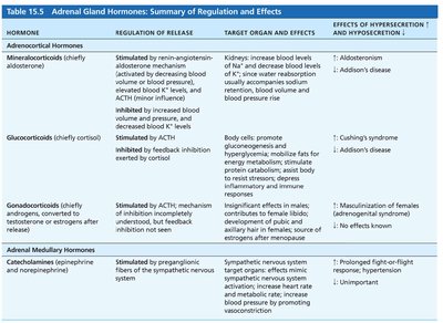
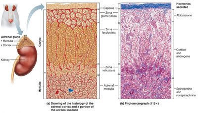
Adrenal Glands
Structure and Hormones
The adrenal glands are paired organs atop the kidneys, consisting of an outer cortex and inner medulla. The cortex produces corticosteroids: mineralocorticoids (aldosterone), glucocorticoids (cortisol), and gonadocorticoids (androgens). The medulla produces catecholamines (epinephrine and norepinephrine).
Mineralocorticoids: Aldosterone
Aldosterone regulates sodium and potassium balance, affecting blood volume and pressure. Its secretion is controlled by the renin-angiotensin-aldosterone mechanism, plasma potassium, ACTH, and atrial natriuretic peptide (ANP).
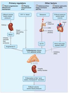
Disorders: Aldosteronism
Hypersecretion of aldosterone (aldosteronism) causes hypertension, edema, and hypokalemia, leading to muscle and neural dysfunction.
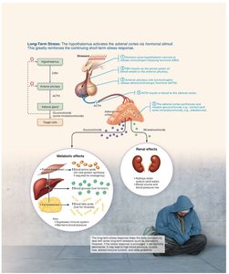
Glucocorticoids: Cortisol
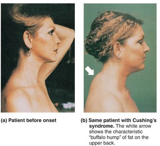
Cortisol helps the body resist stress by increasing blood glucose, fatty acids, and amino acids. It is released in response to ACTH and regulated by negative feedback. Chronic excess leads to Cushing's syndrome, while deficiency causes Addison's disease.

Gonadocorticoids
These weak androgens are converted to testosterone or estrogens in tissues. They contribute to puberty, secondary sex characteristics, and female libido. Hypersecretion can cause masculinization in females and precocious puberty in boys.
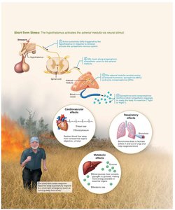
Adrenal Medulla: Catecholamines
The adrenal medulla secretes epinephrine and norepinephrine, which mediate the fight-or-flight response: increasing heart rate, blood pressure, and blood glucose, and diverting blood to essential organs. Effects are short-lived compared to cortical hormones.
Summary Table: Adrenal Gland Hormones
Hormone | Regulation of Release | Target Organ and Effects | Effects of Hyper/Hyposecretion |
|---|---|---|---|
Mineralocorticoids (aldosterone) | Renin-angiotensin, K+, ACTH, ANP | Kidneys: Na+ reabsorption, K+ excretion | ↑: Aldosteronism; ↓: Addison's disease |
Glucocorticoids (cortisol) | ACTH | Body cells: metabolism, stress resistance | ↑: Cushing's; ↓: Addison's |
Gonadocorticoids | ACTH | Sex characteristics, libido | ↑: Masculinization; ↓: No effect |
Catecholamines | Sympathetic nervous system | Fight-or-flight response | ↑: Prolonged response; ↓: Unimportant |