 Back
BackThe Lymphatic System and Body Defenses: Structure, Function, and Immune Response
Study Guide - Smart Notes
 Tailored notes based on your materials, expanded with key definitions, examples, and context.
The Lymphatic System
Overview and Functions
The lymphatic system is a vital component of the circulatory and immune systems, consisting of lymphatic vessels and lymphoid tissues/organs. It is responsible for transporting fluids that have escaped from the cardiovascular system back to the blood and plays essential roles in body defense and resistance to disease.
Lymphatic vessels collect and return excess tissue fluid (lymph) to the bloodstream.
Lymphoid tissues and organs include lymph nodes, spleen, thymus, tonsils, Peyer’s patches, and the appendix.
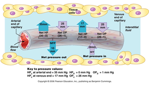
Failure to collect excess fluid results in edema (swelling due to fluid accumulation).
Lymphatic Vessels: Structure and Flow
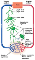
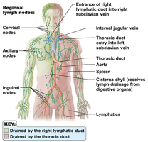
Lymphatic vessels form a one-way system that begins with lymphatic capillaries and flows only toward the heart. These vessels are structurally similar to veins but are thinner-walled, have more valves, and operate under low pressure without a pump.
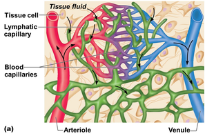
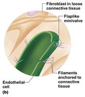
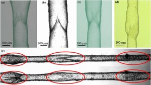
Lymphatic capillaries weave between tissue cells and blood capillaries, with overlapping walls forming flaplike minivalves that allow fluid entry.
Capillaries are anchored to connective tissue by filaments, and higher internal pressure closes the minivalves, forcing fluid along the vessel.
Lymphatic collecting vessels collect lymph from capillaries and return it to veins near the heart via the right lymphatic duct (drains right arm, right side of head and thorax) and thoracic duct (drains the rest of the body).
Lymph transport is aided by skeletal muscle contraction, thoracic pressure changes during breathing, and smooth muscle in vessel walls.
Lymph Nodes and Other Lymphoid Organs
Lymph Nodes: Structure and Function
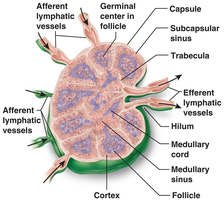
Lymph nodes filter lymph before it is returned to the blood, removing harmful materials such as bacteria, viruses, cancer cells, and foreign substances. Most lymph nodes are kidney-shaped, less than 1 inch long, and buried in connective tissue.
Surrounded by a capsule and divided into compartments by trabeculae.
Cortex (outer part): contains follicles with lymphocytes; germinal centers enlarge during antibody release.
Medulla (inner part): contains phagocytic macrophages.
Defense cells: Macrophages engulf and destroy pathogens; Lymphocytes respond to foreign substances.
Lymph enters via afferent vessels, flows through sinuses, and exits via fewer efferent vessels, slowing flow for filtration.
Other Lymphoid Organs
Spleen: Located on the left side of the abdomen; provides immune surveillance, destroys worn-out red blood cells, filters blood, and stores platelets.
Thymus: Overlies the heart; functions at peak during youth; necessary for T-lymphocyte development and produces thymosin.
Tonsils: Masses of lymphoid tissue around the pharynx; trap and remove pathogens entering the throat.
Peyer’s patches and Appendix: Found in the small intestine and large intestine, respectively; contain macrophages that destroy bacteria.
Mucosa-associated lymphoid tissue (MALT): Includes Peyer’s patches, tonsils, and appendix; protects respiratory and digestive tracts from foreign matter.
Body Defenses: Innate and Adaptive Immunity
Innate (Nonspecific) Defense System
The innate defense system responds immediately to all foreign materials and includes surface barriers and internal defenses.
Surface membrane barriers (first line of defense): skin, mucous membranes, and their secretions (acidic secretions, mucus, gastric juice, lysozyme in saliva and tears).
Cellular and chemical defenses (second line of defense): leukocytes, natural killer cells, phagocytes, inflammatory response, antimicrobial proteins, and fever.
Phagocytosis
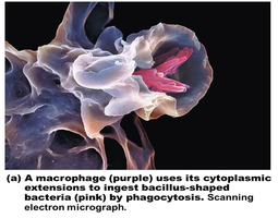
Phagocytes such as neutrophils and macrophages engulf and destroy foreign material. The process involves the phagocyte surrounding the pathogen, internalizing it in a vesicle, and digesting it with lysosomal enzymes.
Inflammatory Response
Triggered by tissue injury; characterized by redness, heat, pain, and swelling.
Inflammatory chemicals (histamine, kinin) cause vasodilation, increased capillary permeability, and attract phagocytes (positive chemotaxis).
Functions: brings in phagocytes, walls off damaged area, disposes of debris/pathogens, and sets the stage for repair.
Antimicrobial Proteins
Complement proteins: At least 20 plasma proteins that, when activated, form membrane attack complexes (MACs) to lyse pathogens and enhance inflammation.
Interferons: Proteins secreted by virus-infected cells that protect neighboring cells from viral infection.
Fever
Systemic response to infection; hypothalamus resets body temperature higher in response to pyrogens.
High temperature inhibits bacterial growth and speeds up repair processes.
Adaptive (Specific) Defense System
The adaptive defense system provides specific resistance to disease through the immune response, targeting antigens with antibodies and specialized cells. It is characterized by antigen specificity, systemic response, and memory.
Humoral immunity: Antibody-mediated; B cells produce antibodies in body fluids.
Cellular immunity: Cell-mediated; T cells target infected or abnormal cells directly.
Antigens and Self-Antigens
Antigens: Substances capable of provoking an immune response (e.g., foreign proteins, nucleic acids, carbohydrates, lipids, pollen, microorganisms).
Self-antigens: Body’s own molecules, usually not antigenic to self but can be antigenic to others (important in transplants and blood compatibility).
Haptens: Incomplete antigens that become antigenic when combined with body proteins (e.g., found in poison ivy, animal dander).
Cells of the Adaptive Defense System
Lymphocytes: B cells (produce antibodies, humoral immunity) and T cells (cell-mediated immunity); develop immunocompetence in bone marrow (B cells) or thymus (T cells).
Antigen-presenting cells (APCs): Dendritic cells, macrophages, and B cells that present antigens to T cells to activate them.
Humoral (Antibody-Mediated) Immune Response
B cells bind to specific antigens, undergo clonal selection, and produce plasma cells (secrete antibodies) and memory cells (provide immunological memory).
Active immunity: Antibodies produced after exposure to antigen (natural infection or vaccination).
Passive immunity: Antibodies obtained from another source (maternal antibodies, immune serum); no immunological memory.
Antibody Structure and Classes
Antibodies (immunoglobulins, Igs) are Y-shaped proteins with variable regions (antigen-binding sites) and constant regions (determine class).
Five major classes: IgM, IgA, IgD, IgG, IgE (each with distinct roles).
Antibody Functions
Inactivate antigens by complement fixation, opsonization, neutralization, agglutination, and precipitation.
Cellular (Cell-Mediated) Immune Response
T cells are activated by antigen presentation (APCs) and require double recognition (antigen and self-protein).
Effector T cells include helper T cells (stimulate other immune cells), cytotoxic T cells (kill infected/abnormal cells), regulatory T cells (suppress immune response), and memory cells (long-term immunity).
Disorders of Immunity
Allergies (Hypersensitivities)
Abnormal, vigorous immune responses to harmless antigens.
Immediate (acute) hypersensitivity: mediated by IgE and histamine (e.g., hives, anaphylaxis).
Delayed hypersensitivity: mediated by T cells and macrophages (e.g., contact dermatitis).
Autoimmune Diseases
Immune system attacks self-tissues due to breakdown of self-tolerance.
Examples: rheumatoid arthritis, myasthenia gravis, multiple sclerosis, Graves’ disease, type 1 diabetes, lupus, glomerulonephritis.
Immunodeficiencies
Congenital or acquired defects in immune function (e.g., SCID, AIDS).
Result from abnormalities in immune cells or complement proteins.
Developmental Aspects of the Lymphatic System and Body Defenses
Lymphatic vessels form from veins during embryonic development; lymph nodes present by the fifth week.
Thymus and spleen are first lymphoid organs to appear; other organs develop later.
Immune response develops around birth; efficiency declines with age, increasing susceptibility to infection, cancer, and autoimmune diseases.
