 Back
BackTissue: The Living Fabric – Study Notes
Study Guide - Smart Notes
 Tailored notes based on your materials, expanded with key definitions, examples, and context.
Tissue: The Living Fabric
Introduction to Tissues
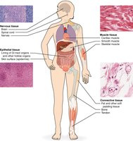
Tissues are groups of cells that are similar in structure and function, forming the basic fabric of the human body. There are four primary tissue types: epithelial, connective, muscle, and nervous tissue. Each type plays a distinct role in maintaining the body's structure and function.
Epithelial Tissue
Characteristics of Epithelial Tissue
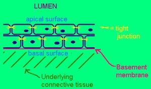
Epithelial tissue covers body surfaces, lines cavities, and forms glands. It is characterized by:
Cellularity: Composed almost entirely of closely packed cells.
Special contacts: Cells are joined by specialized junctions (e.g., tight junctions, desmosomes).
Polarity: Has an apical (free) surface and a basal surface attached to a basement membrane.
Supported by connective tissue: The basal surface is attached to underlying connective tissue.
Avascular but innervated: Contains no blood vessels but is supplied by nerve fibers.
Regenerative: High capacity for renewal and repair.
Locations and Functions of Epithelia
Epithelial tissues are found covering surfaces, lining internal cavities, and composing glands. Their main functions include:
Protection: Shields underlying tissues from mechanical and chemical stress.
Absorption: Uptake of substances (e.g., nutrients in the intestine).
Filtration: Selective movement of substances (e.g., in kidneys).
Excretion: Removal of waste products.
Secretion: Production and release of substances (e.g., mucus, hormones).
Sensory reception: Detects changes in the environment (e.g., skin receptors).
Classification of Epithelia
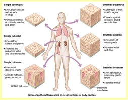
Epithelia are classified by the number of cell layers and the shape of the cells:
Simple: One cell layer thick.
Stratified: Multiple layers of cells.
Squamous: Flat, scale-like cells.
Cuboidal: Cube-shaped cells.
Columnar: Tall, column-like cells.
Special types: Pseudostratified (appears layered but is not), Transitional (changes shape).
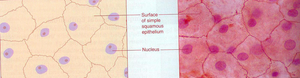
Simple Squamous Epithelium
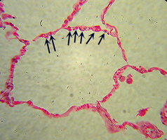
Simple squamous epithelium consists of a single layer of flat cells, resembling a row of fried eggs. It is found in areas where diffusion and filtration occur, such as the inner lining of blood vessels and the alveoli of the lungs.
Functions: Diffusion, filtration
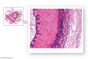
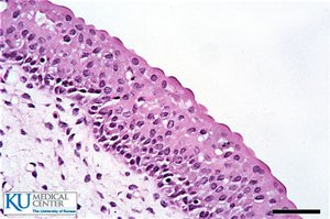
Stratified Squamous Epithelium
This tissue type is composed of many layers of flat cells, providing protection in areas subject to abrasion. It is found in the epidermis, vagina, mouth, and esophagus.
Functions: Protection against mechanical stress and pathogens
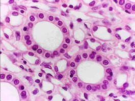
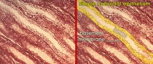
Simple Cuboidal Epithelium
Simple cuboidal epithelium consists of a single layer of cube-shaped cells with central nuclei. It is commonly found in glands, ducts, and kidney tubules.
Functions: Secretion and absorption
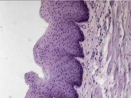
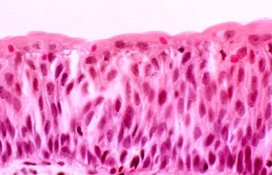
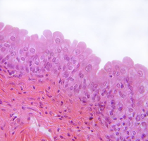
Transitional Epithelium
Transitional epithelium is composed of poufy, dome-shaped cells that can stretch and change shape. It is found in the urinary system, particularly the bladder, allowing for expansion as the bladder fills.
Function: Expansion and recoil
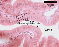
Simple Columnar Epithelium
Simple columnar epithelium consists of tall cells with oval nuclei aligned near the basal surface. It lines much of the digestive tract and is specialized for secretion and absorption.
Functions: Secretion and absorption
Pseudostratified Epithelium
Pseudostratified epithelium appears to have multiple layers due to varying cell heights, but all cells touch the basement membrane. It often contains cilia and goblet cells and is found in the trachea, where it functions in protection and secretion.
Functions: Protection, secretion
Epithelia: Glandular
Glandular epithelia form glands, which are one or more cells that make and secrete an aqueous fluid. Glands are classified by:
Site of product release: Endocrine (into blood) or exocrine (onto surfaces or into ducts)
Number of cells: Unicellular or multicellular
Multicellular Exocrine Glands
These glands are classified by duct type (simple or compound), secretory unit shape (alveolar or tubular), and mode of secretion:
Merocrine: Secretion by exocytosis
Holocrine: Secretion by cell rupture
Epithelial Membranes
Epithelial membranes cover surfaces, line body cavities, and form sheets around organs. They serve to protect and lubricate. There are three main types:
Cutaneous membrane: The skin; a dry membrane containing keratin
Mucous membranes (mucosae): Line body cavities open to the exterior; secrete mucus; found in the digestive, respiratory, and urogenital tracts
Serous membranes (serosae): Line closed ventral body cavities; secrete serous fluid; include pleura, pericardium, and peritoneum
Connective Tissue
Types of Connective Tissue
Connective tissue is the most abundant and widely distributed tissue type. It includes:
Connective tissue proper: Fibroblasts
Cartilage: Chondroblasts
Bone (osseous): Osteoblasts
Blood (liquid): Hematopoietic stem cells
The suffix "-blast" indicates an immature, actively secreting cell; "-cyte" indicates a mature cell.
Characteristics of Connective Tissue
Origin: All connective tissues arise from mesenchyme (embryonic tissue).
Degree of vascularity: Varies from avascular (cartilage) to highly vascular (bone).
Matrix: Extracellular matrix composed of ground substance and fibers, which separates the living cells.
Matrix Components
Ground substance: Interstitial fluid, adhesion proteins, proteoglycans
Fibers: Collagen (strength), elastic (flexibility), reticular (support)
Functions of Connective Tissue
Binds structures together
Supports and protects organs
Hematopoiesis (blood cell formation)
Cushions and insulates
Transports substances (e.g., blood)
Examples of Connective Tissue Types
Areolar: Wraps and cushions organs; found under epithelial tissues
Adipose: Stores fuel, insulates, supports; found under skin, around organs
Hyaline cartilage: Supports, reinforces, resists compression; found in nose, trachea, costal cartilage
Elastic cartilage: Maintains shape with flexibility; found in ear, epiglottis
Fibrocartilage: Absorbs shock; found in intervertebral discs, pubic symphysis, menisci
Bone: Supports, protects, stores minerals; skeleton
Blood: Transports substances; within blood vessels
Connective Tissue Membranes
These membranes are composed solely of connective tissue and include synovial membranes (joints), meninges (brain and spinal cord), fascia, periosteum (bone), perichondrium (cartilage), and pericardium (heart).
Nervous Tissue
Types and Functions
Nervous tissue consists of neurons and neuroglia. Neurons are specialized for transmitting electrical signals, while neuroglia support and protect neurons. Nervous tissue is found in the brain, spinal cord, and nerves, and is responsible for communication and control within the body.
Muscle Tissue
Characteristics and Types
Muscle tissue is specialized for contraction, enabling movement. It is characterized by contractility, extensibility, elasticity, and is well vascularized. Muscle cells are often referred to as "fibers." There are three types:
Skeletal muscle: Voluntary, striated, multinucleated; attached to skeleton; produces movement
Cardiac muscle: Involuntary, striated, branching cells with intercalated discs; found in heart wall; moves blood
Smooth muscle: Involuntary, non-striated; found in walls of hollow organs; moves substances through the body
Tissue Trauma and Repair
Inflammation
Tissue trauma triggers inflammation, characterized by capillary dilation and increased vessel permeability. The classic signs are redness, heat, swelling, and pain.
Steps in Tissue Repair
Inflammation and clot formation
Organization and restoration of blood supply
Regeneration and fibrosis (scar formation)
