 Back
BackLEC 15 Allosteric Regulation and Oxygen-Binding Proteins: Mechanisms and Models
Study Guide - Smart Notes
 Tailored notes based on your materials, expanded with key definitions, examples, and context.
Allosteric Regulation of Proteins
Introduction to Allosteric Regulation
Allosteric regulation is a fundamental mechanism by which the activity of proteins, especially enzymes, is modulated by the binding of effectors at sites distinct from the active site. These effectors induce conformational changes that alter the protein's activity, allowing for fine control of metabolic pathways.
Allosteric proteins are typically oligomeric, meaning they consist of multiple subunits.
Effectors can be activators or inhibitors, and their binding can increase or decrease the protein's affinity for its substrate.
The term "allosteric" derives from Greek: allos (other) and stereos (shape), reflecting the conformational changes induced by effectors.
Models of Allosteric Regulation
The MWC (Monod-Wyman-Changeux) Model
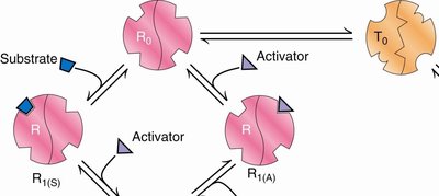
The MWC model, also known as the concerted or symmetry model, describes allosteric proteins as existing in an equilibrium between two states: the T (tense) state and the R (relaxed) state. All subunits are in the same conformation at any given time, and ligand binding shifts the equilibrium between these states.
T state: Low affinity for substrate; predominates in the absence of substrate.
R state: High affinity for substrate; favored upon substrate or activator binding.
Allosteric activators bind preferentially to the R state, shifting the equilibrium toward R and increasing substrate affinity.
Allosteric inhibitors bind preferentially to the T state, shifting the equilibrium toward T and decreasing substrate affinity.
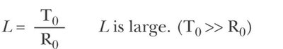
The equilibrium constant for the T to R transition is given by:
where and are the concentrations of the T and R states, respectively. Typically, is large, indicating that the T state predominates in the absence of ligand.
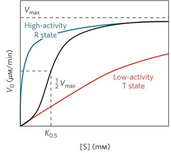
Substrate-Activity Curves in Allosteric Enzymes
Allosteric enzymes display characteristic substrate-activity curves. The presence of activators or inhibitors alters the shape of these curves, reflecting changes in substrate affinity and enzyme activity.
K0.5 is the substrate concentration at half-maximal velocity.
Curves can be hyperbolic (R state), sigmoidal (T state), or a hybrid depending on the state distribution.
Types of Effectors
Positive homotropic effector: The substrate itself enhances its own binding (cooperativity).
Positive heterotropic effector (allosteric activator): A different ligand increases substrate binding.
Negative heterotropic effector (allosteric inhibitor): A different ligand decreases substrate binding.
Allosteric Activators and Inhibitors in the MWC Model
Activators and inhibitors bind to different conformational states, shifting the equilibrium and altering the number of available substrate binding sites.
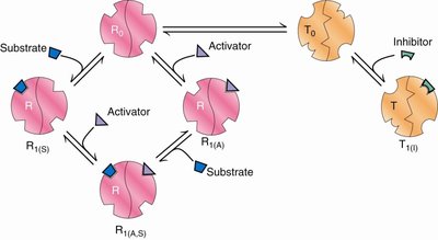
Activators increase the number of substrate binding sites and affinity for substrate.
Inhibitors decrease the number of substrate binding sites and affinity for substrate.
Substrate-Activity Curves with Activators and Inhibitors
Allosteric activators make the curve more hyperbolic, while inhibitors make it more sigmoidal.
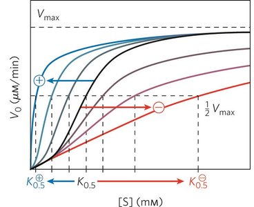
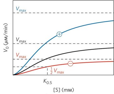
K Systems and V Systems
K system: Effectors change K0.5 (substrate affinity) but not Vmax.
V system: Effectors change Vmax but not K0.5; less common.
Assumptions of the MWC Model
There is a pre-existing equilibrium between R and T states in the absence of ligand.
All subunits are in the same conformation (concerted change).
Ligand binding shifts the equilibrium toward the R state.
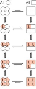
The KNF (Koshland-Némethy-Filmer) Model
The KNF model, or sequential model, proposes that ligand binding induces a conformational change in each subunit individually, which can then influence neighboring subunits. This model can explain both positive and negative cooperativity.
Subunits can exist in different conformations simultaneously.
Ligand binding is sequential and induces conformational changes in adjacent subunits.
Explains both positive and negative cooperativity.
Oxygen-Binding Proteins: Myoglobin and Hemoglobin
Structure and Function
Myoglobin and hemoglobin are classic examples of proteins that exhibit allosteric regulation and cooperativity in ligand binding.
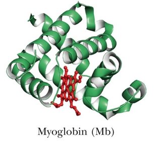
Myoglobin (Mb): Monomeric, oxygen storage protein in muscle.
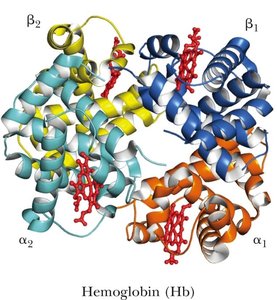
Hemoglobin (Hb): Tetrameric, oxygen transport protein in blood (2 α and 2 β subunits).
Each subunit contains a heme group, a prosthetic group with an iron atom that binds oxygen.
Heme and Oxygen Binding
The heme group consists of a protoporphyrin IX ring with a central Fe2+ ion. Oxygen binding occurs at the iron atom, which is coordinated by histidine residues from the protein.
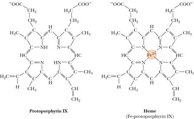
Fe2+ binds O2 reversibly; oxidation to Fe3+ (metmyoglobin) prevents O2 binding.
O2 binding causes a small movement of the Fe2+ into the plane of the porphyrin ring, inducing conformational changes.
Oxygen Binding Curves
Myoglobin displays a hyperbolic O2 binding curve, while hemoglobin displays a sigmoidal curve, indicative of cooperative binding.
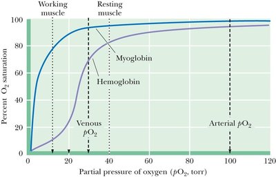
Hemoglobin's sigmoidal curve allows for efficient oxygen loading in the lungs and unloading in tissues.
Mechanism of Cooperativity in Hemoglobin
Oxygen binding to one subunit of hemoglobin increases the affinity of the remaining subunits for oxygen (positive cooperativity). This is achieved through conformational changes transmitted across subunit interfaces.
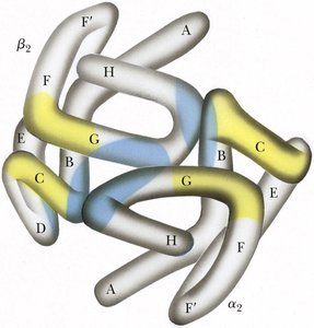
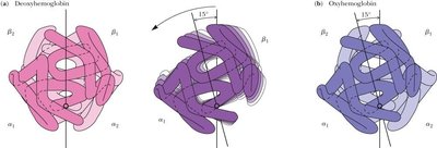
Binding of O2 causes a 15° rotation of one αβ dimer relative to the other, breaking salt bridges and hydrogen bonds.
Bohr Effect and Allosteric Modulation
The Bohr effect describes how H+ and CO2 promote the release of O2 from hemoglobin in tissues. Protonation of specific residues stabilizes the T state, decreasing O2 affinity.
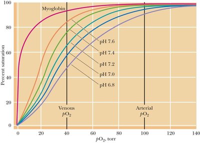
At lower pH (higher [H+]), O2 is released more readily.
CO2 also stabilizes the T state by forming carbamate groups with the N-termini of hemoglobin subunits.
2,3-Bisphosphoglycerate (BPG) Regulation
BPG is a negative allosteric effector that binds between the β subunits of hemoglobin, stabilizing the T state and promoting O2 release in tissues.
BPG is produced during glycolysis and is crucial for hemoglobin function under physiological conditions.
Summary Table: Comparison of Myoglobin and Hemoglobin
Property | Myoglobin | Hemoglobin |
|---|---|---|
Structure | Monomeric | Tetrameric (α2β2) |
Function | Oxygen storage | Oxygen transport |
O2 Binding Curve | Hyperbolic | Sigmoidal (cooperative) |
Allosteric Regulation | None | Yes (H+, CO2, BPG) |
Key Equations
Equilibrium between T and R states:
Oxygen binding (fractional saturation): (for myoglobin)
Recap Questions
A substrate that enhances its own binding is a homotropic modulator.
Allosteric enzymes have their activity changed by changes in intersubunit interactions.
In models of cooperativity, the T state is low affinity and the R state is high affinity.
