 Back
BackAmino Acids, Peptides, and Proteins: Structure, Properties, and Purification
Study Guide - Smart Notes
 Tailored notes based on your materials, expanded with key definitions, examples, and context.
Module 3: Amino Acids, Peptides, and Proteins
Learning Goals
Describe the structures and functions of the 20 common amino acids.
List the names of the 20 common amino acids, including their three- and one-letter codes.
Use experimental data to determine the composition and primary structure of a polypeptide or protein.
Plan a purification to separate a mixture of proteins or amino acids.
Use electrophoresis data to determine the components of a mixture of proteins or amino acids.
Amino Acids
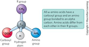
General Structure of Amino Acids
All 20 common amino acids found in proteins are α-amino acids. Each contains a central (α) carbon atom bonded to:
A carboxyl group (–COOH)
An amino group (–NH2)
A hydrogen atom
A distinctive side chain (R group) that determines the amino acid's identity and properties
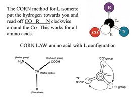
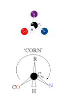
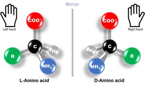
Stereochemistry: L- and D-Forms
Amino acids (except glycine) are chiral and exist as two enantiomers: L and D forms. Only the L-form is found in proteins.
Enantiomers: Stereoisomers that are nonsuperimposable mirror images.
The CORN rule helps distinguish L- from D-amino acids: with the hydrogen atom in front, if the sequence CO–R–N is clockwise, it is the L-form; counterclockwise is the D-form.
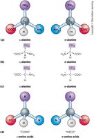
Fischer Projections
Fischer projections are a method to depict the three-dimensional structure of amino acids. Horizontal lines project out of the page, and vertical lines project behind the page. The configuration (L or D) can be determined by tracing the groups around the chiral center.
Amino Acid Shorthand
Each amino acid is identified by:
Full name (e.g., Phenylalanine)
Three-letter code (e.g., Phe)
One-letter symbol (e.g., F)
Classification of Amino Acids by R Group
Amino acids are grouped into five main categories based on the properties of their R groups:
Nonpolar, aliphatic R groups
Aromatic R groups
Polar, uncharged R groups
Positively charged (basic) R groups
Negatively charged (acidic) R groups
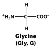
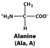
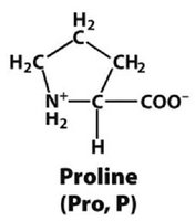
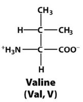
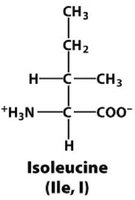
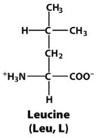
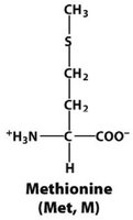
Nonpolar, Aliphatic R Groups
Hydrophobic; often found in the interior of proteins.
Examples: Glycine (Gly, G), Alanine (Ala, A), Proline (Pro, P), Valine (Val, V), Leucine (Leu, L), Isoleucine (Ile, I), Methionine (Met, M)
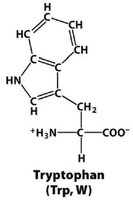
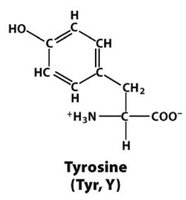
Aromatic R Groups
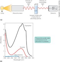
Hydrophobic; absorb ultraviolet light.
Examples: Phenylalanine (Phe, F), Tyrosine (Tyr, Y), Tryptophan (Trp, W)
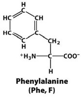
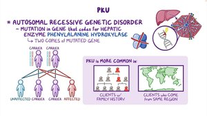
Phenylketonuria (PKU) is a genetic disorder where phenylalanine cannot be metabolized to tyrosine.
Ultraviolet Absorption by Aromatic Amino Acids
Tryptophan and tyrosine, and to a lesser extent phenylalanine, absorb UV light. This property is used to identify and quantify proteins.

Polar, Uncharged R Groups
Hydrophilic; can form hydrogen bonds.
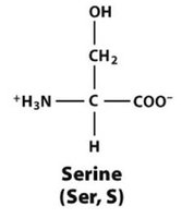
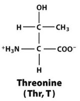
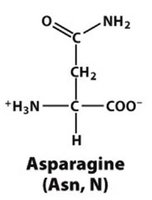
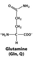
Examples: Serine (Ser, S), Threonine (Thr, T), Asparagine (Asn, N), Glutamine (Gln, Q), Cysteine (Cys, C), Tyrosine (Tyr, Y)
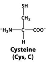
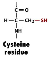
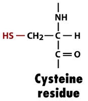
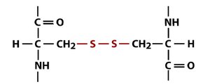
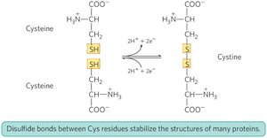
Disulfide Bonds
Cysteine residues can form disulfide bonds (–S–S–) upon oxidation, stabilizing protein structure.
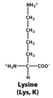
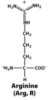
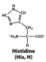
Positively Charged (Basic) R Groups
Hydrophilic; basic side chains.
Examples: Lysine (Lys, K), Arginine (Arg, R), Histidine (His, H)
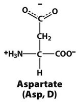
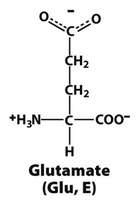
Negatively Charged (Acidic) R Groups
Hydrophilic; acidic side chains.
Examples: Aspartate (Asp, D), Glutamate (Glu, E)
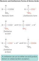
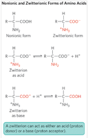
Acid-Base Properties of Amino Acids
Amino acids can act as both acids and bases (amphoteric). The pKa value indicates the tendency to lose a proton. At physiological pH, amino acids exist as zwitterions (dipolar ions with both positive and negative charges).
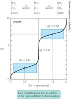
Titration Curves
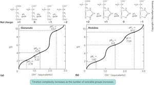
The titration curve of an amino acid shows how its charge changes with pH. Each ionizable group has a characteristic pKa. Amino acids with ionizable R groups have more complex titration curves.
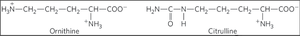
Uncommon Amino Acids
In addition to the 20 common amino acids, many uncommon amino acids exist and play important roles in metabolism and specialized functions (e.g., ornithine, citrulline).

Peptides and Proteins
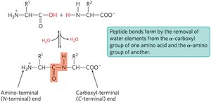
Peptide Bonds
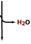
Proteins are polymers of amino acids linked by peptide bonds, which are amide linkages formed by the condensation of the α-carboxyl group of one amino acid and the α-amino group of another, releasing water.
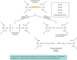
Disulfide Bonds in Proteins
Disulfide bonds between cysteine residues stabilize protein structure by forming covalent links within or between polypeptide chains.
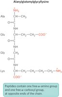
Ionization Behavior of Peptides
In a polypeptide, only the terminal α-amino and α-carboxyl groups are free and ionizable; all others are involved in peptide bonds. Ionizable side chains contribute to the overall charge and properties of the peptide.
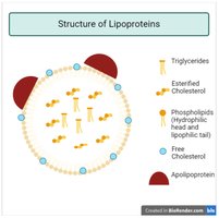
Conjugated Proteins
Some proteins contain non-amino acid components, such as lipids (lipoproteins), carbohydrates (glycoproteins), or metal ions (metalloproteins). These are called conjugated proteins.
Purifying Proteins
Principles of Protein Purification
To study proteins, they must be purified from complex mixtures. Common separation methods include chromatography (based on size, charge, or binding properties) and electrophoresis.
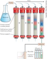
Detection and Quantification of Proteins
Proteins are detected and quantified based on their specific functions. For enzymes, activity (amount of substrate converted per unit time) and specific activity (enzyme units per mg protein) are key measures.
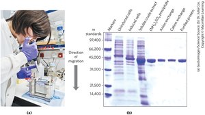
Electrophoresis
Electrophoresis separates proteins based on their charge-to-mass ratio and is used to estimate the number of proteins in a mixture and assess purity.
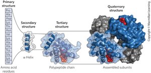
The Primary Structure of Proteins and Protein Chemistry
Levels of Protein Structure
Primary structure: Linear sequence of amino acids in a polypeptide chain (covalent bonds).
Secondary structure: Local folding into structures such as α-helices and β-sheets (hydrogen bonds).
Tertiary structure: Overall three-dimensional folding of a single polypeptide chain.
Quaternary structure: Arrangement of multiple polypeptide subunits.
Protein Function and Sequence
The function of a protein is determined by its amino acid sequence. Proteins with similar functions often have similar sequences, and sequence comparison provides evolutionary insights.
Fragmentation and Sequencing
Polypeptide chains can be fragmented by chemical or enzymatic methods for sequencing. Mass spectrometry is a powerful tool for determining molecular mass, sequence, and analyzing entire proteomes.
Consensus Sequences
Consensus sequences highlight conserved regions among related proteins, indicating functional or structural importance.
Summary
The 20 common amino acids share a general structure but differ in their R groups, which determine their properties and classification.
Amino acids can act as acids and bases, exist as zwitterions, and have characteristic titration curves.
Proteins are polymers of amino acids linked by peptide bonds, with structure and function determined by their sequence.
Protein purification and analysis rely on techniques such as chromatography and electrophoresis.
Knowledge of amino acid sequences provides biochemical and evolutionary information.
