 Back
BackAmino Acids, Peptides, and Proteins: Structure, Properties, and Function
Study Guide - Smart Notes
 Tailored notes based on your materials, expanded with key definitions, examples, and context.
Amino Acids, Peptides, and Proteins
Overview of Protein Functions
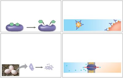
Proteins are essential macromolecules that perform a vast array of functions in biological systems. Their diversity in structure allows them to catalyze reactions, provide structural support, transport molecules, and more.
Enzymatic proteins: Catalyze biochemical reactions, e.g., digestive enzymes hydrolyze food molecules.
Defensive proteins: Protect against disease, e.g., antibodies neutralize pathogens.
Storage proteins: Store amino acids, e.g., casein in milk, ovalbumin in eggs.
Transport proteins: Move substances, e.g., hemoglobin transports oxygen.
Hormonal proteins: Coordinate physiological processes, e.g., insulin regulates blood sugar.
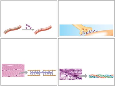
Receptor proteins: Detect chemical signals, e.g., nerve cell receptors.
Contractile and motor proteins: Enable movement, e.g., actin and myosin in muscles.
Structural proteins: Provide support, e.g., keratin in hair, collagen in connective tissue.
Amino Acids: Building Blocks of Proteins
General Structure and Properties
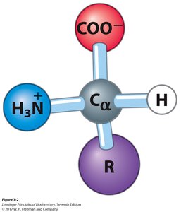
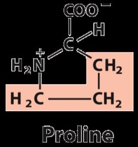
Amino acids are organic molecules with a central (α) carbon atom bonded to an amino group, a carboxyl group, a hydrogen atom, and a variable side chain (R group). The properties of the R group determine the characteristics and classification of each amino acid.
Polymerization: Amino acids link via peptide bonds to form proteins.
Acid-base properties: Amino acids can act as buffers due to their ionizable groups.
Physical and chemical diversity: The 20 standard amino acids have varied side chains, conferring diverse properties.
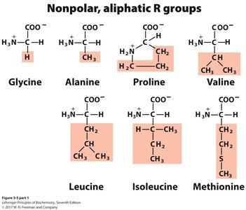
Classification of Amino Acids
Amino acids are classified based on the properties of their side chains (R groups):
Nonpolar, aliphatic: Glycine, Alanine, Proline, Valine, Leucine, Isoleucine, Methionine
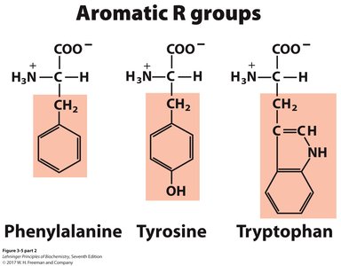
Aromatic: Phenylalanine, Tyrosine, Tryptophan
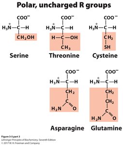
Polar, uncharged: Serine, Threonine, Cysteine, Asparagine, Glutamine
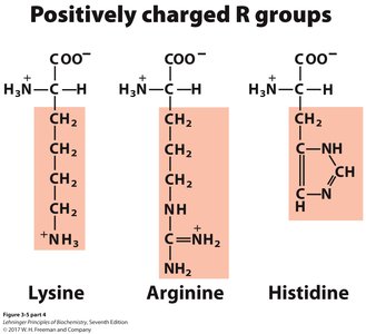
Positively charged (basic): Lysine, Arginine, Histidine
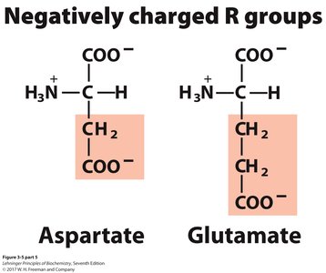
Negatively charged (acidic): Aspartate, Glutamate
Properties and Conventions of Amino Acids
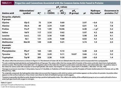
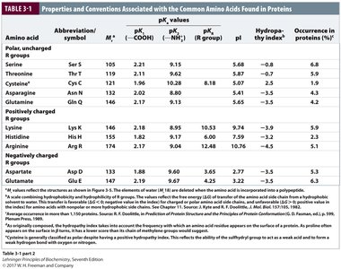
The physical and chemical properties of amino acids, such as molecular weight, pKa values, isoelectric point (pI), and hydropathy index, are summarized in the following tables:
Amino Acid | Abbreviation | pK1 (COOH) | pK2 (NH3+) | pKR | pI | Hydropathy Index | Occurrence (%) |
|---|---|---|---|---|---|---|---|
Glycine | Gly, G | 2.34 | 9.60 | - | 5.97 | -0.4 | 7.2 |
Alanine | Ala, A | 2.34 | 9.69 | - | 6.01 | 1.8 | 7.8 |
Serine | Ser, S | 2.21 | 9.15 | - | 5.68 | -0.8 | 6.8 |
Lysine | Lys, K | 2.18 | 8.95 | 10.53 | 9.74 | -3.9 | 5.9 |
Aspartate | Asp, D | 1.88 | 9.60 | 3.65 | 2.77 | -3.5 | 5.3 |
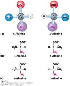
Chirality of Amino Acids
All amino acids except glycine are chiral, existing as L- or D-isomers. Proteins are composed exclusively of L-amino acids. The configuration can be determined using the 'CoRN' rule.
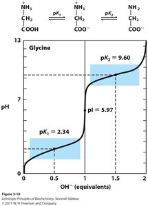
Ionization and Acid-Base Properties of Amino Acids
Ionization Behavior
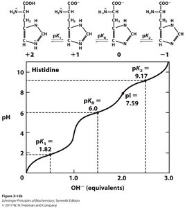
Amino acids contain at least two ionizable groups: the α-carboxyl and α-amino groups, each with characteristic pKa values. The ionization state depends on the pH of the environment.
At low pH, amino acids are fully protonated (cationic form).
At high pH, they are fully deprotonated (anionic form).
At intermediate pH, they exist as zwitterions (net charge zero).
The isoelectric point (pI) is the pH at which the net charge is zero:
Amino acids with ionizable side chains have a third pKa and more complex titration curves.
Isoelectric Point Calculation Example
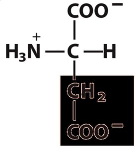
For amino acids with ionizable side chains, the pI is calculated by averaging the two pKa values that surround the zwitterionic form. For example, aspartate:
Peptides and Proteins: Structure and Hierarchy
Peptide Bond Formation
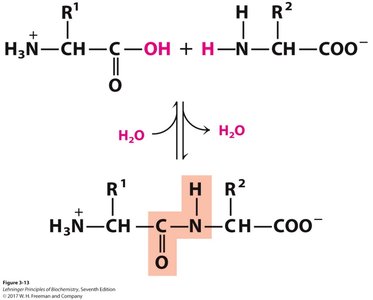
Peptides are formed by condensation reactions between amino acids, creating amide (peptide) bonds. The sequence is always written from the N-terminus to the C-terminus.
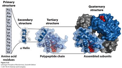
Levels of Protein Structure
Primary structure: Linear sequence of amino acids.
Secondary structure: Local folding into α-helices and β-sheets, stabilized by hydrogen bonds.
Tertiary structure: Overall 3D shape of a single polypeptide, stabilized by hydrophobic interactions, hydrogen bonds, ionic interactions, and disulfide bridges.
Quaternary structure: Assembly of multiple polypeptide subunits into a functional protein complex.
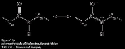
Peptide Bond Properties
The peptide bond is planar due to resonance, which restricts rotation and contributes to the rigidity of the protein backbone. However, rotation is possible around the bonds adjacent to the α-carbon, allowing for the formation of secondary structures.
Secondary Structures: α-Helix and β-Sheet
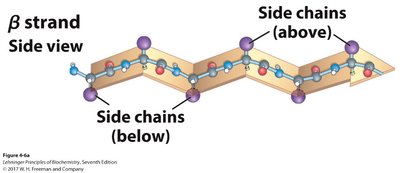
The α-helix is a right-handed coil stabilized by hydrogen bonds between every fourth amino acid. The β-sheet consists of extended strands connected laterally by hydrogen bonds, which can be parallel or antiparallel.
Tertiary and Quaternary Structure
Tertiary structure is the overall 3D arrangement of a polypeptide, while quaternary structure refers to the assembly of multiple polypeptides. These structures are stabilized by hydrophobic interactions, hydrogen bonds, ionic interactions, and sometimes covalent disulfide bonds.
Fibrous Proteins: Structure and Function
Examples of Fibrous Proteins
Structure | Characteristics | Examples |
|---|---|---|
α-Helix, cross-linked by disulfide bonds | Tough, insoluble, protective | α-Keratin (hair, feathers, nails) |
β-Conformation | Soft, flexible filaments | Silk fibroin |
Collagen triple helix | High tensile strength, no stretch | Collagen (tendons, bone matrix) |
Protein Purification and Analysis
Separation Techniques
Proteins can be separated and purified based on their physical and chemical properties, such as charge (ion exchange chromatography), size (size exclusion chromatography), affinity for ligands (affinity chromatography), solubility, and hydrophobicity.
Protein Sequencing
Edman degradation: Classical method for sequencing by stepwise removal of N-terminal residues.
Mass spectrometry: Modern method for determining peptide mass and sequence, including post-translational modifications.
Summary
Proteins are polymers of amino acids with diverse structures and functions.
Amino acids are classified by their side chains and have characteristic acid-base properties.
Protein structure is hierarchical: primary, secondary, tertiary, and quaternary levels.
Fibrous proteins have specialized structural roles, while globular proteins perform dynamic functions.
Protein purification and sequencing are essential for biochemical analysis.
