 Back
BackBacterial Cell Structure and Cell Wall Infection Science: Study Notes
Study Guide - Smart Notes
 Tailored notes based on your materials, expanded with key definitions, examples, and context.
Bacterial Cell Structure
Core Components of Bacterial Cells
Bacterial cells are characterized by a simple yet efficient design, consisting of essential structural and functional elements. These components are critical for cellular processes, survival, and adaptation.
Chromosome: Typically a single, circular DNA molecule that governs cell function and genetic inheritance.
Plasmid: Small, circular DNA molecules carrying accessory genes, often conferring antibiotic resistance.
Ribosome: Protein synthesis machinery, composed of RNA and protein.
Cytoplasm: Gel-like matrix where metabolic reactions occur.
Cell Wall: Provides structural integrity and protection.
Capsule: An external polysaccharide layer aiding in immune evasion and surface adherence.
Fimbriae: Short, hair-like structures for attachment to surfaces.
Flagellum: Long, whip-like appendage for motility.
Example: Escherichia coli possesses all these components, enabling survival in diverse environments. 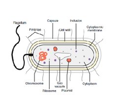
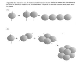
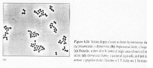
Bacterial Shapes and Arrangements
Cocci and Bacilli Arrangements
Bacteria exhibit distinct shapes and arrangements, which are genetically determined and influence their identification and pathogenicity.
Cocci: Spherical bacteria; arrangements include diplococcus (pairs), chains, tetrads, packets, and clusters.
Bacilli: Rod-shaped bacteria; arrangements include single bacillus, diplobacilli, streptobacilli, and coccobacillus.
Spiral Forms: Includes vibrio (comma-shaped), spirillum (rigid spiral), and spirochete (flexible coil).
Example: Staphylococcus aureus forms clusters, while Bacillus subtilis forms chains. 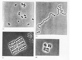
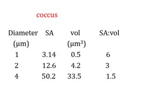
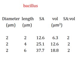
Surface Area to Volume Ratio
The surface area to volume (SA:vol) ratio affects nutrient uptake and metabolic efficiency.
Coccus: As diameter increases, SA:vol decreases.
Bacillus: Length increases with constant diameter, SA:vol remains relatively stable.
Example: Smaller cells have higher SA:vol, facilitating faster exchange of materials.
Diameter (μm) | SA | vol (μm³) | SA:vol |
|---|---|---|---|
1 | 3.14 | 0.5 | 6 |
2 | 12.6 | 4.2 | 3 |
4 | 50.2 | 33.5 | 1.5 |
Diameter (μm) | length (μm) | SA | vol (μm³) | SA:vol |
|---|---|---|---|---|
2 | 2 | 12.6 | 6.3 | 2 |
2 | 4 | 25.1 | 12.6 | 2 |
2 | 6 | 37.7 | 18.8 | 2 |
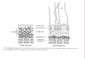
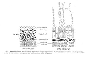
Bacterial Cell Wall Structure
Peptidoglycan and Structural Polysaccharides
The bacterial cell wall is primarily composed of peptidoglycan, a structural polysaccharide that provides rigidity and protection.
Peptidoglycan: Consists of repeating units of N-acetylglucosamine (GlcNAc) and N-acetylmuramic acid (MurNAc) linked by peptide chains.
Cellulose: β-glucose chains, found in plant cell walls.
Chitin: N-acetylglucosamine, found in fungal walls and insect exoskeletons.
Lysozyme: Enzyme that cleaves MurNAc–GlcNAc bonds, weakening the cell wall.
Example: Peptidoglycan is targeted by antibiotics such as penicillin.
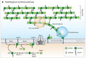
Peptidoglycan Synthesis Pathway
Peptidoglycan synthesis involves multiple enzymatic steps, both in the cytoplasm and at the cell membrane.
Cytoplasm: MurA–MurF enzymes build UDP-linked precursors.
Membrane: MraY and MurG attach precursors to undecaprenyl phosphate (Und-P), forming Lipid I and II.
Transport: MurJ flips Lipid II to the extracellular side.
Extracellular: PG polymerase adds glycan strands; PG cross-linker links peptides for wall strength.
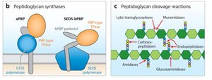
Peptidoglycan Enzymes
Enzymes involved in peptidoglycan synthesis and cleavage are essential for cell wall remodeling and division.
Synthesis: aPBP complex (GT51 polymerase + PBP transpeptidase), SEDS-bPBP complex (SEDS polymerase + PBP + pedestal).
Cleavage: Muramidase, transglycosylase, carboxy/endopeptidase, amidase, glucosaminidase.
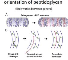
Peptidoglycan Enlargement (Gram-Positive)
Gram-positive bacteria expand their cell wall by a mechanical process involving cleavage, insertion, and re-linking of peptidoglycan strands.
Cross-link cleavage: Old bonds are cut.
Nascent glycan strand insertion: New strands are added.
Cross-link formation: New bonds stabilize the wall.
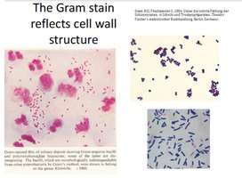
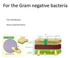
Gram Stain and Cell Wall Structure
The Gram stain differentiates bacteria based on cell wall structure:
Gram-positive: Thick peptidoglycan layer, retains crystal violet stain (purple).
Gram-negative: Thin peptidoglycan layer, outer membrane, loses crystal violet and stains pink with safranin.
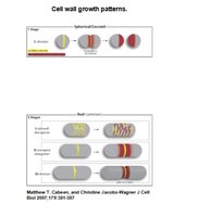
Cell Wall Growth Patterns
Cell wall growth varies by shape:
Spherical (cocci): Growth occurs at multiple sites.
Rod-shaped (bacilli): Growth occurs along the length or at the poles.
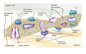
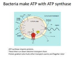
Bacterial Membrane Structure and Transport
Membrane Structure and Function
Bacterial membranes are composed of a phospholipid bilayer with embedded proteins, functioning as a selective barrier and facilitating transport.
Fluid Mosaic Model: Membrane proteins and lipids move laterally, allowing flexibility.
Barrier Function: Controls entry and exit of ions, solutes, and metabolites.
Transport Mechanisms:
Simple diffusion: Small molecules (O2, CO2, NH3).
Facilitated diffusion: Water via aquaporins.
Passive transport: Charged ions via ion channels.
Active transport: Larger compounds (nutrients).
Bacterial Chromosomes and Plasmids
Chromosome Structure
Bacterial chromosomes are double-stranded DNA, compacted into the nucleoid region.
Supercoiling: DNA is tightly packed, often 1000x the length of the cell.
Multiple Copies: Some bacteria have more than one chromosome copy.
Plasmids
Plasmids are extrachromosomal DNA elements that facilitate genetic movement and confer 'luxury' functions such as antibiotic resistance.
Horizontal Gene Transfer (HGT): Acquisition of new DNA via conjugation, transformation, or transduction.
Bacterial Survival: Endospores
Endospore Formation
Endospores are highly resistant structures produced by certain bacteria (e.g., Bacillus and Clostridium species) to survive extreme conditions.
Sporulation: Process of endospore formation when conditions are unfavorable.
Resistance: Endospores withstand heat, desiccation, chemicals, and radiation.
 Example: Bacillus anthracis and Clostridium tetani produce endospores, enabling persistence in harsh environments.
Additional info: Some explanations and tables were expanded for academic completeness and clarity.
