 Back
BackBiochem Midterm study guide slightly messed up.
Study Guide - Smart Notes
 Tailored notes based on your materials, expanded with key definitions, examples, and context.
Carbohydrates: Structure and Function
Role of Carbohydrates
Carbohydrates are essential biomolecules involved in energy generation, storage, molecular recognition, cellular protection, cell signaling, adhesion, lubrication, protein trafficking, and maintenance of biological structure.
Energy: Carbohydrates serve as primary energy sources and storage molecules.
Molecular Recognition: Glycans on cell surfaces mediate recognition and signaling.
Structural Roles: Polysaccharides like cellulose and chitin provide structural integrity.
Molecular Basis and Structure of Carbohydrates
Carbohydrates are carbon-rich molecules with multiple hydroxyl (-OH) groups. Their basic formula is (C-H2O)n, reflecting their nature as carbon hydrates.
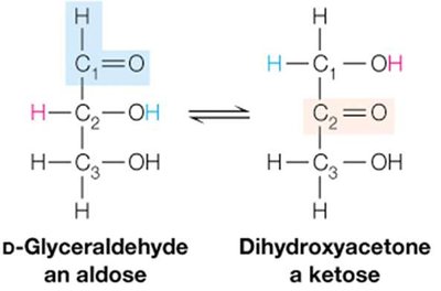
Monosaccharides: The simplest carbohydrates, classified as aldoses (with an aldehyde group) or ketoses (with a ketone group).
Polymeric Chains: Monosaccharides link to form oligosaccharides and polysaccharides.
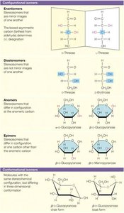
Isomerism in Carbohydrates
Carbohydrates exhibit various forms of isomerism, including tautomers, enantiomers, diastereomers, anomers, and epimers.
Tautomers: Molecules with the same formula but different connectivity, interconvertible under catalysis.
Enantiomers: Chiral molecules that are nonsuperimposable mirror images (D and L forms).
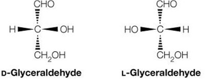
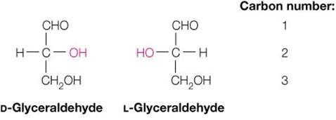
D-monosaccharides: Most common in nature; L forms have specialized roles.
Diastereomers: Isomers with multiple chiral centers that are not mirror images.
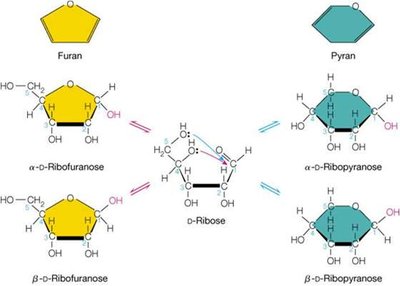
Cyclic Forms and Anomers
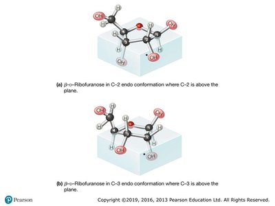
Monosaccharides with five or more carbons form cyclic structures (furanose and pyranose rings) via internal hemiacetal formation. Anomers differ at the carbonyl carbon (C1), designated as α or β based on the position of the hydroxyl group.
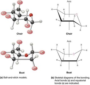
Conformational Isomers
Pyranose rings can adopt chair or boat conformations, with the chair being more stable due to minimized steric strain.
Configurational Isomers: Anomers, Epimers, Diastereomers
Configurational isomers require breaking and reforming covalent bonds to interconvert. Anomers differ at the anomeric carbon, epimers differ at one chiral center, and diastereomers differ at multiple centers.
Monosaccharide Modifications
Monosaccharides can be modified by reactions with alcohols, amines, and phosphates, increasing their biochemical versatility for signaling and metabolic functions.


Sugar phosphate esters: Important metabolic intermediates.
Lactones and acids: Oxidized forms of monosaccharides.
Alditols: Reduced forms, e.g., glucose to sorbitol.
Amino sugars: Amino acid derivatives of sugars.
Glycosides: Formed by elimination of water between the anomeric hydroxyl and another compound.
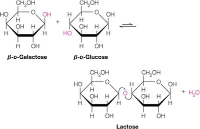
Oligosaccharides and Disaccharides
Monosaccharides form glycosidic bonds to create oligosaccharides and disaccharides, which serve as energy stores and intermediates.
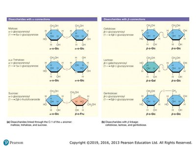
Glycosyltransferases: Enzymes catalyzing glycosidic bond formation.
Disaccharide Features: Defined by monomer identity, linkage position, order, and anomeric configuration.
Polysaccharides
Polysaccharides serve as energy storage (starch, glycogen) and structural materials (cellulose, chitin). They can be homopolysaccharides (one monomer type) or heteropolysaccharides (two or more types).
Storage: Amylose, amylopectin (plants), glycogen (animals).
Structure: Cellulose (plants), chitin (organisms), glycosaminoglycans (vertebrates).
Glycoproteins
Proteins with covalently attached oligo- or polysaccharide chains, important for cell recognition and signaling.
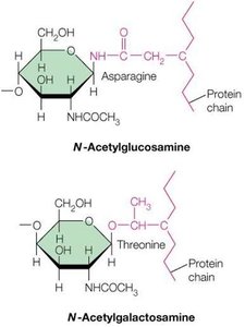
N-linked: Attached via N-acetylglucosamine to asparagine.
O-linked: Attached via N-acetylgalactosamine to threonine or serine.
Lipids, Membranes, and Cellular Transport
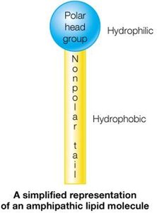
Lipid Structure and Function
Lipids are hydrophobic, water-insoluble molecules that serve as energy stores, membrane components, and signaling molecules. Unlike other biomolecules, lipids associate via noncovalent interactions.
Classes of Lipids
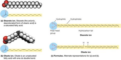
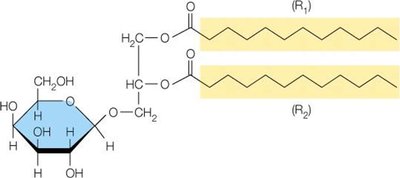
Free Fatty Acids: Simplest lipids, with hydrophilic carboxylate and hydrophobic hydrocarbon tail. Saturated (no double bonds) and unsaturated (one or more double bonds).
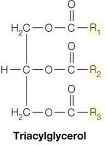
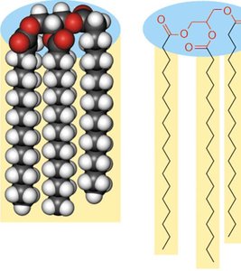
Triacylglycerols: Fat storage molecules, triesters of fatty acids and glycerol.
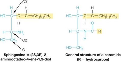
Phospholipids: Major membrane-forming lipids, with diverse headgroups.
Glycolipids: Lipids with carbohydrate groups.
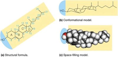
Steroids: Lipids with fused ring structures, e.g., cholesterol.
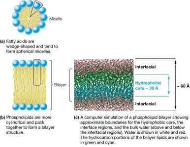
Membrane Structure and Fluid Mosaic Model
Biological membranes are sheet-like structures composed of lipids and proteins, forming barriers and mediating cellular functions. Membranes are asymmetric, fluid, and electrically polarized.
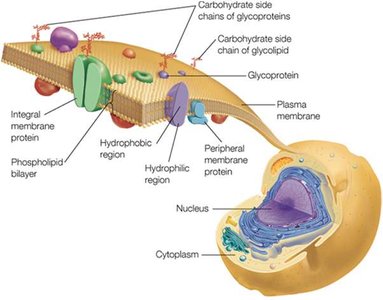
Peripheral Proteins: Exposed on one side.
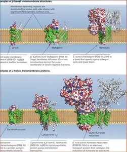
Integral Proteins: Span the membrane, involved in transport and signaling.
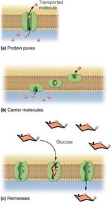
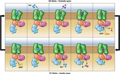
Transport Across Membranes
Membranes are selectively permeable, with transport mediated by proteins. Transport mechanisms include diffusion, facilitated transport, passive and active transport, ion channels, and pumps.
Mechanisms of Signal Transduction
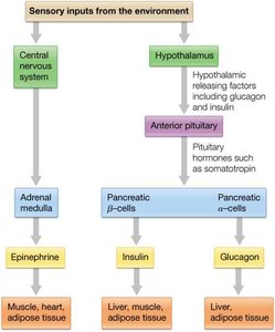
Hormones and Receptors
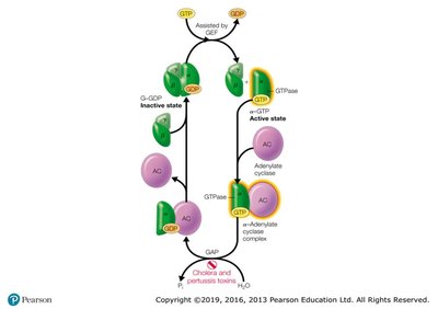
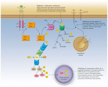
Hormones are signaling molecules (peptides, steroids, amino acid derivatives) that regulate enzyme activity, gene expression, and membrane permeability. Membrane-bound receptors mediate signal transduction via second messengers and ion channels.
Protein Function and Structure
Antibodies and Immunoglobulins
Antibodies are large proteins with variable domains for specific antigen binding, crucial for immune response. They consist of heavy and light chains, held by disulfide bonds.
Globins: Oxygen Binding Proteins
Myoglobin and hemoglobin are oxygen-binding proteins with heme prosthetic groups. Hemoglobin exhibits cooperative binding and allosteric effects.
Motor Proteins: Actin and Myosin
Motor proteins convert ATP hydrolysis into mechanical work, essential for muscle contraction and cellular motility.
Enzymes: Biological Catalysts
Enzyme Structure and Function
Enzymes are highly specific catalysts, accelerating reactions by stabilizing the transition state and lowering activation energy. They require cofactors (coenzymes or metals) for activity.
Free Energy and Reaction Kinetics
Spontaneity of reactions is determined by free energy change (ΔG). Enzymes do not affect ΔG but increase reaction rates by lowering activation energy (ΔG≠).
Enzyme-Substrate Complex and Models
Substrates bind to the enzyme's active site, forming an enzyme-substrate (ES) complex. Two models describe this interaction: lock and key (enzyme fits substrate) and induced fit (enzyme changes shape upon binding).
Enzyme Kinetics and Regulation
Michaelis-Menten Kinetics: Describes the rate of enzymatic reactions.
Enzyme Modulation: Influenced by temperature, pH, and inhibitors (competitive, uncompetitive, noncompetitive, irreversible).
Allosteric Enzymes: Respond to environmental signals and utilize feedback controls.
Nucleic Acids: Structure and Function
DNA and RNA
Nucleic acids are informational macromolecules composed of nucleotide monomers. DNA stores genetic information; RNA facilitates protein synthesis and gene regulation.
Monomer Structure: 5-carbon sugar, nucleobase, phosphate group.
Polymerization: Phosphodiester bonds link monomers, forming the backbone.
Nucleobases: Purines (A, G), Pyrimidines (C, T/U).
Primary, Secondary, and Tertiary Structure
Primary: Linear sequence of nucleotides.
Secondary: Double helix, base pairing, antiparallel strands.
Tertiary: Supercoiling, chromosomal organization.
Central Dogma
Replication: DNA copying.
Transcription: DNA to RNA.
Translation: RNA to protein.
Biochemistry Foundations
Major Classes of Biomolecules
Proteins: Structure, function, catalysis.
Nucleic Acids: Information storage and transfer.
Carbohydrates: Energy and cell communication.
Lipids: Membranes, energy storage, signaling.
Chemical Bonds and Water
Covalent Bonds: Strong, stable, electron sharing.
Noncovalent Bonds: Weaker, dynamic, include charge-charge, dipole, van der Waals, hydrogen bonds.
Water: Excellent solvent, hydrogen bonding, high surface tension, hydrophilic/hydrophobic effects.
Acids, Bases, and pH
Acid: Proton donor.
Base: Proton acceptor.
pH: Measure of H+ concentration.
Henderson-Hasselbalch Equation:
Bioenergetics and Thermodynamics
First Law: Energy conservation.
Second Law: Entropy increases.
Free Energy:
Spontaneity: Negative ΔG is favorable.
Protein Structure
Primary: Amino acid sequence.
Secondary: α-helix, β-sheet.
Tertiary: 3D folding.
Quaternary: Multi-subunit complexes.
Amino Acids
Structure: α-carbon, amino group, carboxyl group, side chain (R).
Properties: Nonpolar, polar, charged, essential/nonessential.
Peptide Bond: Amide linkage, planar, cis/trans forms.
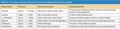
Summary Table: Disaccharides
Disaccharide | Structure | Natural Occurrence | Physiological Role |
|---|---|---|---|
Sucrose | Glc(α1→2)Fru | Many fruits, seeds, roots, honey | Primary energy source in many organisms |
Lactose | Gal(β1→4)Glc | Milk, some plant sources | Major animal energy source |
Trehalose | Glc(α1→1)Glc | Insects, other animals, insect blood | Major circulatory sugar in insects |
Maltose | Glc(α1→4)Glc | Plants (starch) and animals (glycogen) | Intermediate in starch/glycogen metabolism |
Cellobiose | Glc(β1→4)Glc | Plants (cellulose) | Dimer of cellulose polymer |
Gentiobiose | Glc(β1→6)Glc | Some plants (e.g., gentian) | Constituent of plant glycosides/polysaccharides |
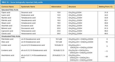
Summary Table: Fatty Acids
Common Name | Systematic Name | Abbreviation | Structure | Melting Point (°C) |
|---|---|---|---|---|
Capric acid | Decanoic acid | 10:0 | CH3(CH2)8COOH | 31.6 |
Palmitic acid | Hexadecanoic acid | 16:0 | CH3(CH2)14COOH | 63.1 |
Oleic acid | cis-9-Octadecenoic acid | 18:1cisΔ9 | CH3(CH2)7CH=CH(CH2)7COOH | 16 |
Linoleic acid | cis,cis-9,12-Octadecadienoic acid | 18:2Δ9,12 | CH3(CH2)7CH=CHCH2CH=CH(CH2)6COOH | -5 |
Additional info: Tables above are reconstructed from lecture content and textbook figures for clarity and completeness.
