 Back
BackNucleotides and Nucleic Acids: Structure, Function, and Metabolism
Study Guide - Smart Notes
 Tailored notes based on your materials, expanded with key definitions, examples, and context.
Nucleotides and Nucleic Acids
Basic Structure of Nucleotides
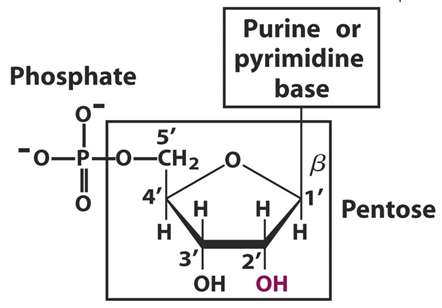
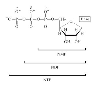
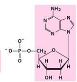
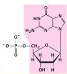
Nucleotides are the fundamental building blocks of nucleic acids, such as DNA and RNA. Each nucleotide consists of three main components: a nitrogenous base, a pentose sugar, and one or more phosphate groups. The absence of the phosphate group yields a nucleoside.
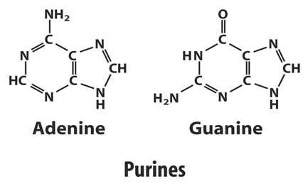
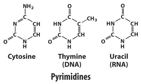
Nitrogenous base: Can be a purine (adenine, guanine) or a pyrimidine (cytosine, thymine, uracil).
Pentose sugar: Either ribose (in RNA) or deoxyribose (in DNA).
Phosphate group: Attached to the 5' carbon of the pentose sugar, forming the backbone of nucleic acids.

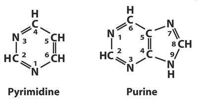
Nitrogenous Bases
Nitrogenous bases are classified into two groups: purines and pyrimidines. These bases are weakly basic, hydrophobic, and relatively insoluble in water at neutral pH. Their solubility increases at acidic or alkaline pH due to ionization.
Purines: Double-ring structures (adenine and guanine).
Pyrimidines: Single-ring structures (cytosine, thymine, and uracil).
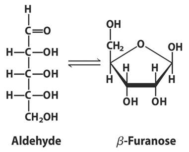
Pentose Sugars
The pentose sugar in nucleotides can exist in a straight-chain (aldehyde or ketone) or cyclic (furanose) form. In aqueous solution, monosaccharides predominantly exist as cyclic molecules due to the formation of a covalent bond between the carbonyl group and a hydroxyl group along the chain.
Phosphate Group and Nucleic Acid Backbone
The phosphate group is esterified to the 5' carbon of the pentose sugar. Together, the pentose and phosphate form the hydrophilic backbone of nucleic acids. The phosphate groups are fully ionized and negatively charged at physiological pH, contributing to the solubility and structural stability of nucleic acids.
Types and Roles of Nucleotides
Major Functions
Constituents of nucleic acids (DNA and RNA)
Energy currency in metabolism (e.g., ATP)
Structural components of enzyme cofactors and metabolic intermediates (e.g., NAD, FAD, CoA)
Essential chemical links to hormones and other extracellular stimuli
Nucleic Acids: DNA and RNA
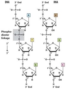
Nucleic acids are polymers of nucleotides. DNA contains deoxyribose, while RNA contains ribose. The sequence of nucleotides is read from the 5' to 3' direction.
Deoxyribonucleotides and Ribonucleotides
Deoxyribonucleotides are the monomers of DNA, while ribonucleotides are the monomers of RNA. The main difference is the presence of a hydroxyl group at the 2' position in ribose (RNA) and its absence in deoxyribose (DNA).
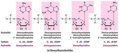
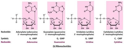
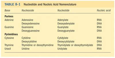
Nucleotide and Nucleic Acid Nomenclature
The nomenclature of nucleotides, nucleosides, and nucleic acids is systematic and distinguishes between the base, nucleoside, and nucleotide forms for both purines and pyrimidines.
Base | Nucleoside | Nucleotide | Nucleic Acid |
|---|---|---|---|
Adenine | Adenosine | Adenyate | RNA |
Deoxyadenosine | Deoxyadenylate | DNA | |
Guanine | Guanosine | Guanylate | RNA |
Deoxyguanosine | Deoxyguanylate | DNA | |
Cytosine | Cytidine | Cytidylate | RNA |
Deoxycytidine | Deoxycytidylate | DNA | |
Thymine | Thymidine or deoxythymidine | Thymidylate or deoxythymidylate | DNA |
Uracil | Uridine | Uridylate | RNA |
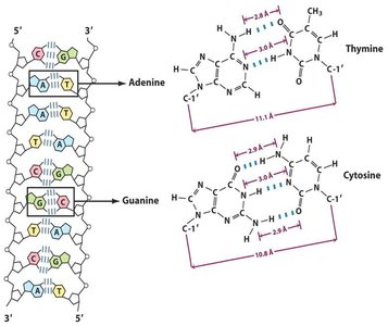
DNA Structure and Base Pairing
Double Helix Structure
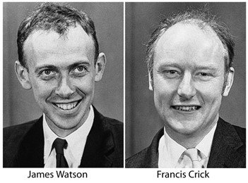
DNA is a double helix, with two antiparallel strands held together by hydrogen bonds between complementary bases. The structure was elucidated by Watson and Crick, with key experimental evidence from Rosalind Franklin and Maurice Wilkins.
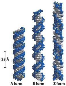
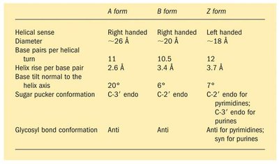
Forms of DNA
DNA can exist in several conformations, including A, B, and Z forms, which differ in helical sense, diameter, base pairs per turn, and other structural features.
A form | B form | Z form | |
|---|---|---|---|
Helical sense | Right handed | Right handed | Left handed |
Diameter | ~26 Å | ~20 Å | ~18 Å |
Base pairs per turn | 11 | 10.5 | 12 |
Helix rise per base pair | 2.6 Å | 3.4 Å | 3.7 Å |
Base tilt normal to helix axis | 20° | 6° | 7° |
Sugar pucker conformation | C-3' endo | C-2' endo | C-2' endo for pyrimidines; C-3' endo for purines |
Glycosyl bond conformation | Anti | Anti | Anti for pyrimidines; syn for purines |
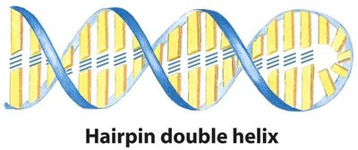
RNA Structure
RNA is typically single-stranded but can form complex secondary structures such as hairpins, bulges, and internal loops due to intramolecular base pairing.
Biological Roles of Nucleotides
Energy Currency: ATP
Adenosine triphosphate (ATP) is the primary energy carrier in cells. The hydrolysis of its phosphoanhydride bonds releases significant free energy, which is used to drive various biochemical reactions.
Ester linkage: ~14 kJ/mol
Phosphoanhydride linkage: ~30 kJ/mol
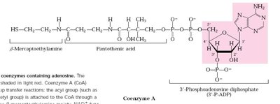
Cofactors and Metabolic Intermediates
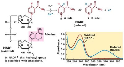
Nucleotides are integral components of several essential cofactors, including Coenzyme A (CoA) and Nicotinamide adenine dinucleotide (NAD+).
Biosynthesis and Degradation of Nucleotides
Pathways of Nucleotide Biosynthesis
Nucleotides are synthesized via two main pathways: de novo and salvage. The de novo pathway constructs nucleotides from simple precursors, while the salvage pathway recycles free bases and nucleosides from nucleic acid breakdown.
De novo pathway: Begins with amino acids, ribose 5-phosphate, CO2, and NH3.
Salvage pathway: Recycles free bases and nucleosides.
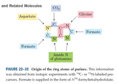
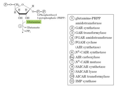
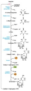
De Novo Synthesis of Purines
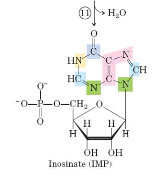
The purine ring is built atom by atom on a ribose phosphate scaffold. The first intermediate with a complete purine ring is inosinate (IMP). The process is regulated by feedback inhibition.
De Novo Synthesis of Pyrimidines
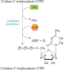
The pyrimidine ring is synthesized as orotate, which is then attached to ribose phosphate. This pathway is also regulated by feedback inhibition.
Degradation of Purines and Pyrimidines
Purine degradation produces uric acid, while pyrimidine degradation produces urea. These processes are essential for nucleotide turnover and nitrogen balance in the cell.
Salvage Pathways
Salvage pathways recover free purine and pyrimidine bases released during nucleotide degradation. One primary reaction is catalyzed by adenosine phosphoribosyltransferase, where free adenine reacts with PRPP to yield AMP and pyrophosphate.
Reaction:

Ribonucleotides as Precursors of Deoxyribonucleotides
Ribonucleotides are converted to deoxyribonucleotides, which are essential for DNA synthesis. This reduction is catalyzed by ribonucleotide reductase and is a key regulatory step in DNA biosynthesis.
Additional info: The reduction of ribonucleotides to deoxyribonucleotides is crucial for maintaining balanced pools of DNA precursors and is tightly regulated to ensure genomic stability.
