 Back
BackProtein Function: Oxygen Binding and Allostery in Hemoglobin and Myoglobin
Study Guide - Smart Notes
 Tailored notes based on your materials, expanded with key definitions, examples, and context.
Protein Function
Introduction to Protein Function
Proteins are essential macromolecules that perform a vast array of functions in biological systems, including catalysis, structure, transport, and regulation. The relationship between protein structure and function is a central theme in biochemistry, as the three-dimensional conformation of a protein determines its specific activity.
Oxygen Transport Proteins: Hemoglobin and Myoglobin
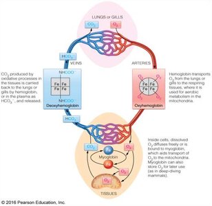
Oxygen is vital for cellular respiration and energy production. Specialized proteins, hemoglobin (Hb) and myoglobin (Mb), facilitate the transport and storage of oxygen in vertebrates. These proteins allow for a significantly greater amount of oxygen to be carried in the blood than would be possible by simple dissolution.
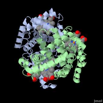
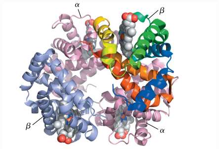
Hemoglobin (Hb): A heterotetrameric protein found in red blood cells, responsible for oxygen transport from lungs to tissues.
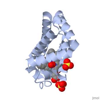
Myoglobin (Mb): A monomeric protein found in muscle tissue, primarily involved in oxygen storage and release during periods of high demand.
Structural Features and Heme Binding
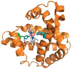
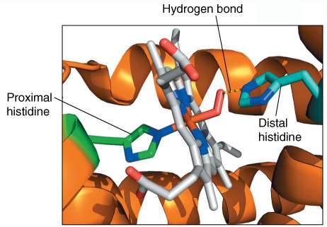
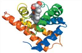
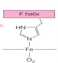
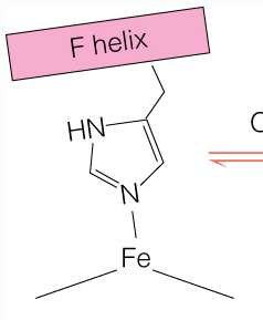
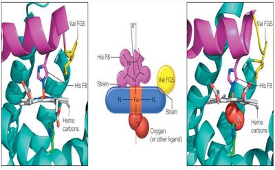
Both Hb and Mb contain a heme prosthetic group, which is responsible for reversible oxygen binding. The heme consists of a protoporphyrin IX ring coordinated to an Fe2+ ion. Key amino acid residues within the binding pocket, such as the proximal and distal histidines, stabilize heme binding and modulate oxygen affinity.
Proximal Histidine: Directly coordinates with Fe2+, anchoring the heme group.
Distal Histidine: Forms a hydrogen bond with bound O2, increasing selectivity for O2 over CO.
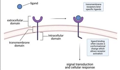
Ligand Binding and Dissociation Constant (KD)
The equilibrium dissociation constant (KD) quantitatively describes the affinity between a protein (receptor) and its ligand. A lower KD value indicates higher affinity. The binding equilibrium can be represented as:
Fractional Occupancy (θ):
At 50% occupancy:
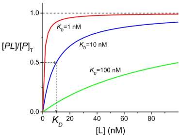
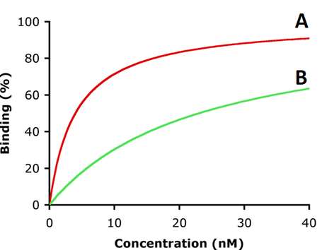
Comparing Protein Affinity
Proteins with lower KD values have higher affinity for their ligands. This can be visualized using binding curves, where the ligand concentration at 50% occupancy (P50 for gases like O2) is analogous to KD.

Oxygen Binding Curves: Myoglobin vs. Hemoglobin
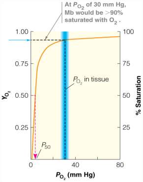
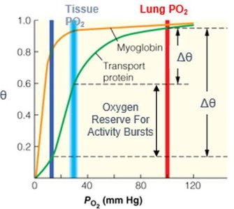
Myoglobin exhibits a hyperbolic binding curve, reflecting its high affinity and single binding site. Hemoglobin, with four subunits, displays a sigmoidal curve due to cooperative binding, allowing efficient oxygen loading in the lungs and unloading in tissues.
Hyperbolic Curve (Mb): High affinity, low KD, efficient storage but poor release.
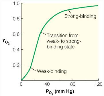
Sigmoidal Curve (Hb): Cooperative binding, affinity changes with O2 concentration, optimal for transport.
Cooperativity and the Hill Equation
Hemoglobin's cooperative binding violates the assumption of independent binding sites. The Hill equation models this behavior:
n (Hill coefficient): Indicates degree of cooperativity (n > 1: positive, n = 1: none, n < 1: negative).
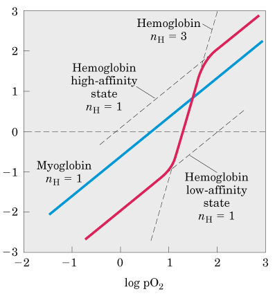
Hill Plot Analysis
The Hill plot linearizes the binding data to determine the Hill coefficient (nH):
Slope: Equals nH, reflecting cooperativity.
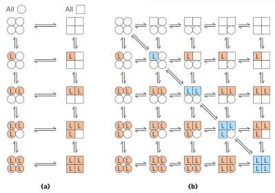
Allosteric Models: MWC and KNF
Two main models describe allosteric transitions in hemoglobin:
Monod-Wyman-Changeux (MWC) Model: All subunits switch between T (tense, low affinity) and R (relaxed, high affinity) states simultaneously (concerted model).
Koshland-Némethy-Filmer (KNF) Model: Subunits change conformation sequentially upon ligand binding (sequential model).
Structural Basis of T to R Transition
Oxygen binding induces a transition from the T-state (low affinity) to the R-state (high affinity) in hemoglobin. This involves conformational changes in the heme group and surrounding protein structure, including rotation of the F-helix and disruption of salt bridges.
T-state: Heme is domed, salt bridges stabilize structure, low O2 affinity.
R-state: Heme is planar, salt bridges broken, high O2 affinity.
The Bohr Effect
The Bohr effect describes the influence of pH and CO2 on hemoglobin's oxygen affinity. Lower pH (higher CO2) decreases affinity, promoting O2 release in metabolically active tissues. This effect is crucial for efficient oxygen delivery during exercise or hypoxia.
High pH (lungs): Higher O2 affinity, O2 loading.
Low pH (tissues): Lower O2 affinity, O2 unloading.
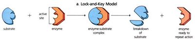
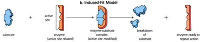
Lock-and-Key vs. Induced-Fit Models
Protein-ligand interactions can be described by two models:
Lock-and-Key Model: Assumes rigid protein and ligand shapes that fit perfectly.
Induced-Fit Model: Protein undergoes conformational change upon ligand binding, optimizing interaction.
Summary Table: Myoglobin vs. Hemoglobin
Property | Myoglobin | Hemoglobin |
|---|---|---|
Structure | Monomer | Tetramer (α2β2) |
O2 Binding Curve | Hyperbolic | Sigmoidal |
Function | O2 storage | O2 transport |
Cooperativity | None | Positive |
Location | Muscle | Blood |
Additional info: The notes above integrate foundational concepts in protein function, ligand binding, and allosteric regulation, with a focus on hemoglobin and myoglobin as model systems. These principles are broadly applicable to other protein-ligand interactions in biochemistry.
