 Back
BackProtein Function: Oxygen Binding and Allostery in Hemoglobin and Myoglobin
Study Guide - Smart Notes
 Tailored notes based on your materials, expanded with key definitions, examples, and context.
Protein Function
Introduction to Protein Function
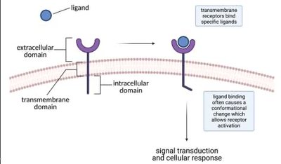
Proteins are essential biomolecules that perform a wide variety of functions in living organisms, including catalysis, transport, signaling, and structural support. The relationship between a protein's structure and its function is a central concept in biochemistry.
Structure-Function Relationship: The three-dimensional structure of a protein determines its specific function.
Example: Oxygen transport and storage are mediated by the proteins hemoglobin and myoglobin, respectively.
Oxygen Transport Proteins: Hemoglobin and Myoglobin
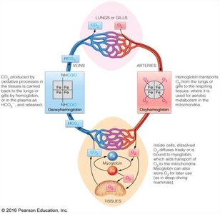
Oxygen is vital for cellular respiration and energy production. Specialized proteins enable efficient oxygen transport and storage in the body.
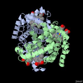
Hemoglobin (Hb): A heterotetrameric protein found in red blood cells, responsible for transporting oxygen from the lungs to tissues.
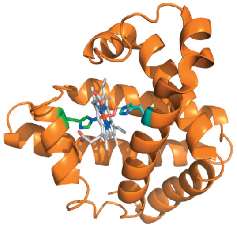
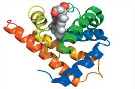
Myoglobin (Mb): A monomeric protein found in muscle tissue, responsible for oxygen storage and release during muscle contraction.
Oxygen Binding: Both proteins bind oxygen via a heme prosthetic group, but differ in their affinity and physiological roles.
Heme Group and Oxygen Binding
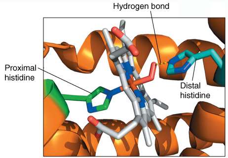
The heme group is a planar, aromatic structure containing an iron (Fe2+) ion at its center, which binds oxygen reversibly.
Protoporphyrin IX: The organic component of heme.
Iron Coordination: The Fe2+ ion forms six coordination bonds: four with nitrogen atoms of the porphyrin ring, one with a proximal histidine residue, and one with oxygen (or another ligand).
Stabilization: Key residues in the binding pocket, such as proximal and distal histidines, stabilize heme and modulate ligand binding.
Ligand Binding and Dissociation Constant (KD)
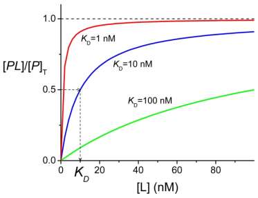
Ligand binding to proteins is described by equilibrium and kinetic parameters. The equilibrium dissociation constant (KD) quantifies the affinity between a protein and its ligand.
Ligand: A molecule that binds specifically to a protein (e.g., O2, CO, substrate, inhibitor).
KD Definition: where [P] is free protein, [L] is free ligand, and [PL] is the protein-ligand complex.
Affinity: Lower KD values indicate higher affinity.
Fractional Occupancy (θ): , where [P]T is total protein concentration.
Relationship to Binding Curve: At θ = 0.5, [L] = KD.
Comparing Protein Affinity
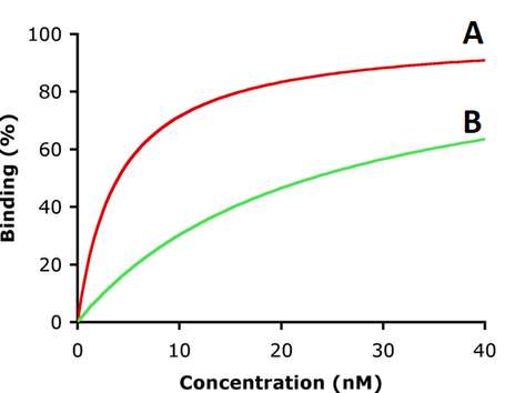
Comparing the KD values of different proteins for the same ligand allows assessment of their relative affinities.
Example: If Protein A has KD = 5 nM and Protein B has KD = 30 nM, Protein A has higher affinity for the ligand.
Oxygen Binding Curves: Myoglobin vs. Hemoglobin
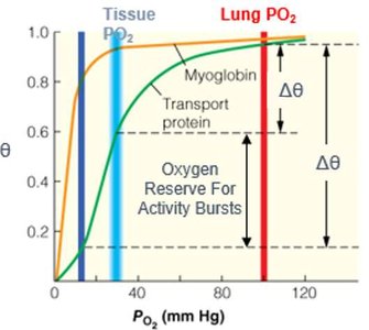
The oxygen binding behavior of myoglobin and hemoglobin can be visualized using binding curves, which plot fractional saturation (θ) against oxygen partial pressure (pO2).
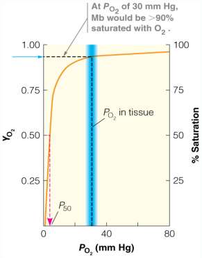
Myoglobin: Displays a hyperbolic binding curve, indicating a single high-affinity binding site.
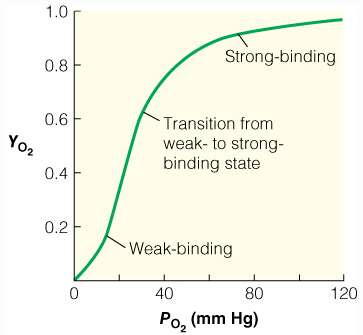
Hemoglobin: Displays a sigmoidal (S-shaped) curve, reflecting cooperative binding among its four subunits.
P50: The partial pressure of O2 at which the protein is 50% saturated; analogous to KD for gases.
Cooperativity and Allosteric Regulation
Hemoglobin exhibits cooperative binding, where the binding of one O2 molecule increases the affinity of the remaining subunits for O2. This is a form of allosteric regulation.
Cooperativity: Positive cooperativity results in a sigmoidal binding curve; negative cooperativity would result in a less steep curve.
Allosteric Effect: Conformational changes in one subunit affect the binding properties of others.
Hill Equation: Used to quantify cooperativity: , where n is the Hill coefficient.
Hill Plot: A linear transformation of the binding curve to determine the degree of cooperativity.
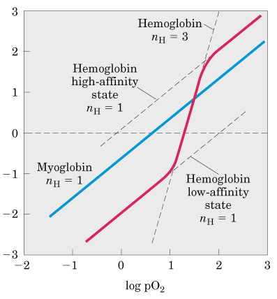
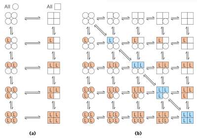
Allosteric Models: MWC and KNF
Two main models describe allosteric transitions in multimeric proteins:
Monod-Wyman-Changeux (MWC) Model: Proposes that all subunits exist in either a tense (T) or relaxed (R) state, and ligand binding shifts the equilibrium between these states in a concerted manner.
Koshland-Némethy-Filmer (KNF) Model: Suggests that subunits change conformation sequentially upon ligand binding, with each binding event increasing the likelihood of neighboring subunits switching to the high-affinity state.
Structural Basis of Cooperativity in Hemoglobin
Hemoglobin's quaternary structure enables communication between subunits, facilitating cooperative binding. The transition from the T (tense, low affinity) to R (relaxed, high affinity) state involves significant conformational changes.
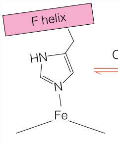
Heme Flattening: O2 binding flattens the heme group, shifting the position of the proximal histidine and the F helix, which propagates structural changes throughout the protein.
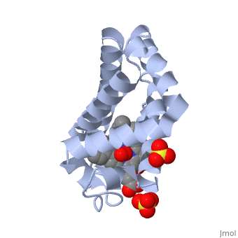
Salt Bridges: Stabilize the T-state; their disruption upon O2 binding favors the R-state.

The Bohr Effect
The Bohr effect describes the influence of pH and CO2 concentration on hemoglobin's oxygen-binding affinity. Lower pH (higher CO2) decreases affinity, promoting O2 release in metabolically active tissues.
Mechanism: Protonation of specific residues stabilizes the T-state, facilitating O2 offloading where it is most needed.
Physiological Relevance: Enhances O2 delivery during exercise or hypoxia.
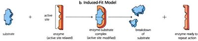
Protein-Ligand Binding Models
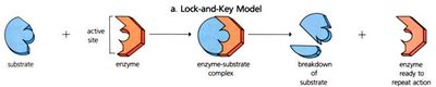
Two classical models describe how proteins interact with their ligands:
Lock-and-Key Model: Assumes a rigid protein structure with a binding site complementary to the ligand.
Induced-Fit Model: Proposes that ligand binding induces a conformational change in the protein, optimizing the interaction.
Summary Table: Comparison of Myoglobin and Hemoglobin
Property | Myoglobin | Hemoglobin |
|---|---|---|
Structure | Monomer | Tetramer (α2β2) |
O2 Binding Curve | Hyperbolic | Sigmoidal |
Cooperativity | None | Positive |
Main Function | O2 storage | O2 transport |
P50 (O2 affinity) | Low (high affinity) | Higher (lower affinity in tissues) |
Additional info: The notes above integrate foundational concepts in protein function, ligand binding, and allosteric regulation, with a focus on hemoglobin and myoglobin as model systems in biochemistry. The Bohr effect and cooperative binding are critical for understanding physiological oxygen delivery.
