 Back
BackTriacylglycerols, Phospholipids, and Sphingolipids: Structure, Function, and Disease
Study Guide - Smart Notes
 Tailored notes based on your materials, expanded with key definitions, examples, and context.
Triacylglycerols and Their Role in Energy Storage
Structure of Triacylglycerols
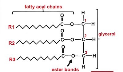
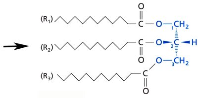
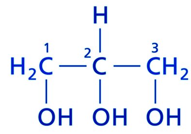
Triacylglycerols (triglycerides) are the primary form of energy storage in many organisms. They consist of a glycerol backbone, which is a three-carbon alcohol, esterified with three fatty acid chains. The fatty acids can vary in length and saturation, contributing to the physical properties of the triacylglycerol.
Glycerol Backbone: Three-carbon molecule with hydroxyl groups at each carbon.
Fatty Acyl Chains: Each hydroxyl group is esterified with a fatty acid, forming three ester bonds.
Hydrophobic Nature: Triacylglycerols are highly hydrophobic, making them suitable for energy storage.
Physical State: The melting point depends on the chain length and degree of unsaturation of the fatty acids.
Functions of Triacylglycerols
Metabolic Energy: Stored in small deposits in cells for metabolic energy.
Long-Term Storage: Accumulated in adipocytes under the skin and abdomen for long-term energy storage.
Thermal Insulation: Provides insulation in aquatic mammals.
Examples of Triacylglycerols
Animal Fat: High concentration of tripalmitin (three palmitic acid chains, 16:0), melting point ~63°C.
Olive Oil: Major component is triolein (three oleic acid chains, 18:1 Δ9), melting point ~13°C.
Butter: Mixture of palmitate, myristate, stearate, oleate, and shorter fatty acids, melting points between 70°C and 13°C.
Glycerophospholipids: Structure and Function
Core Structure and Common Head Groups
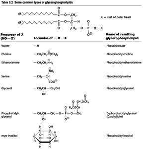
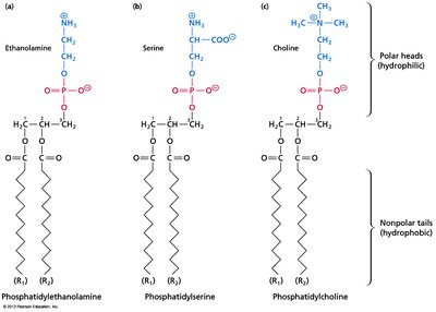
Glycerophospholipids are essential components of cellular membranes. They are based on a glycerol backbone, but differ from triacylglycerols by having two fatty acid chains and a phosphate group at the third position. The phosphate group can be further modified by various head groups, resulting in different types of glycerophospholipids.
R1: Usually a saturated fatty acid.
R2: Usually an unsaturated fatty acid.
Phosphate Group: Present as a diester compound, often with additional head groups.
Amphipathic Nature: Contains a hydrophobic tail and a hydrophilic head.
Membrane Formation
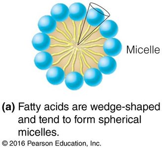
Micelles: Fatty acids form spherical micelles due to their wedge shape.
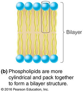
Bilayers: Glycerophospholipids form bilayer structures, which are the basis of biological membranes.
Common Glycerophospholipids
Phosphatidylcholine
Phosphatidylethanolamine
Phosphatidylserine
Phosphatidylglycerol
Phosphatidylinositol
Cardiolipin (Diphosphatidylglycerol)
Phospholipases: Enzymatic Hydrolysis of Phospholipids
Types and Functions
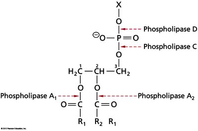
Phospholipases are enzymes that hydrolyze phospholipids at specific positions. They play roles in digestion, cell signaling, and can be found in venoms.
Phospholipase A1: Releases the fatty acid at the first position.
Phospholipase A2: Releases the fatty acid at the second position.
Phospholipase C and D: Cleave at different sites on the phosphate group.
Biological Roles: Digestive enzymes, signal transduction, and cell membrane remodeling.
Plasmalogens: Structure and Function
Structure of Plasmalogens
Plasmalogens are a unique class of glycerophospholipids characterized by a vinyl ether linkage at the C1 position of glycerol, instead of the typical ester linkage. The head group can be ethanolamine or choline.
Vinyl Ether Linkage: Provides distinct chemical properties.
Head Groups: Ethanolamine and choline are common.
Membrane Function: Important for membrane stability and organization, especially in lipid rafts.
Prevalence: ~10% of phospholipids in humans, mostly in membranes.

Sphingolipids: Structure, Classes, and Functions
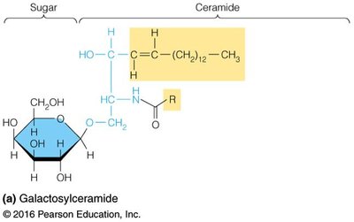
Core Structure: Sphingosine and Ceramide
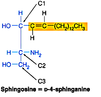
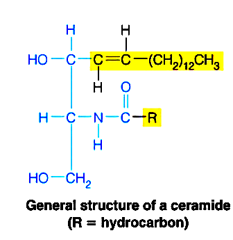
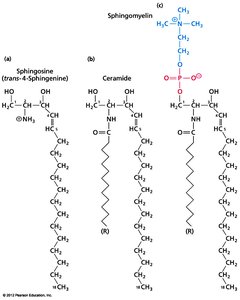
Sphingolipids are the second most abundant lipids in membranes. They are built on the amino alcohol sphingosine, which forms the backbone. Ceramide is formed when a fatty acid is linked to the NH2 group of sphingosine via an amide bond.
Sphingosine: Long-chain amino alcohol.
Ceramide: Sphingosine with a fatty acid attached via an amide bond.
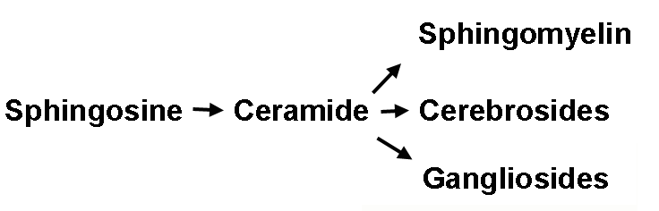
Classes of Sphingolipids
Sphingolipids are classified based on the head group attached to the C1 atom of ceramide. All have two hydrophobic tails for membrane insertion and a polar head group.
Sphingomyelin: Similar to phosphatidylcholine, major component of myelin sheaths.
Cerebrosides: Contain a monosaccharide group, common in brain and nerve cell membranes.
Gangliosides: Contain a complex carbohydrate chain, act as cell surface markers, mostly found in CNS.
Cerebrosides and Gangliosides
Cerebrosides: Sphingolipids with a single sugar group attached to ceramide, e.g., galactocerebrosides in nerve tissue.
Gangliosides: Sphingolipids with a complex carbohydrate chain, including sialic acid, important for cell-cell interaction and recognition.
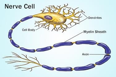
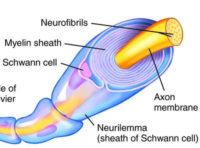
Myelin Sheath and Lipid Composition in Nerve Cells
Role of Sphingolipids in Myelin Sheath
The myelin sheath is a lipid-rich structure that insulates nerve cells and facilitates rapid signal transmission. Sphingomyelin and cerebrosides are major components of myelin.
Function: Protects and insulates nerve cells, essential for proper nervous system function.
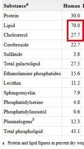
Composition: High lipid content, including cholesterol, sphingomyelin, cerebrosides, and plasmalogens.
Disease Associated with Sphingolipid Degradation
Sphingolipidoses: Genetic diseases caused by deficiencies in enzymes responsible for sphingolipid degradation, leading to accumulation of sphingolipids in tissues.
Examples: Tay-Sachs disease, Gaucher disease, Niemann-Pick disease.
Symptoms: Neurological impairment, organ dysfunction, and developmental delays.
Additional info: Sphingolipids are also involved in cell signaling and recognition, and their dysfunction can impact immune responses and cell communication.
