 Back
BackBio 100 LEC Chapter 6 Part 1
Study Guide - Smart Notes
 Tailored notes based on your materials, expanded with key definitions, examples, and context.
Bio 100 LEC Chapter 6 Part 1
Introduction to Cells
Cells are the fundamental units of life, and all living organisms are composed of cells. The study of cells provides insight into the continuity and complexity of life, as all cells arise from pre-existing cells through division. Understanding cell structure and function is essential for comprehending biological processes at all levels.
Biologists Use Microscopes and Biochemistry to Study Cells
Microscopy: Revealing Cellular Structure
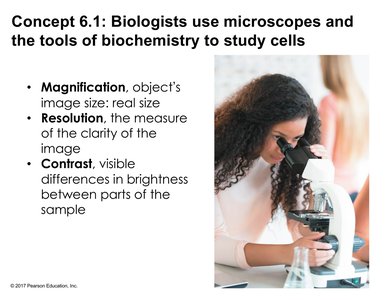
Microscopes are essential tools for visualizing cells and their components, which are often too small to be seen with the unaided eye. Three key properties of microscopy are:
Magnification: The ratio of an object's image size to its real size.
Resolution: The measure of the clarity of the image; the minimum distance two points can be separated and still be distinguished as separate points.
Contrast: The visible differences in brightness between parts of the sample, often enhanced by staining or optical techniques.
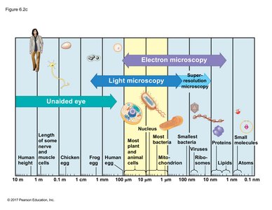
Size Range of Cells and Organelles
The size of cells and their components varies widely, from atoms and small molecules to entire cells. Most cells are between 1 and 100 micrometers in diameter, requiring light or electron microscopy for visualization.
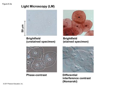
Types of Light Microscopy
Brightfield (unstained specimen): Light passes directly through the specimen; little contrast and detail.
Brightfield (stained specimen): Staining enhances contrast and reveals more detail, such as nuclei and membranes.
Phase-contrast: Enhances contrast in unstained cells by amplifying variations in density.
Differential Interference Contrast (DIC/Nomarski): Uses polarized light to produce images with a 3D-like appearance.
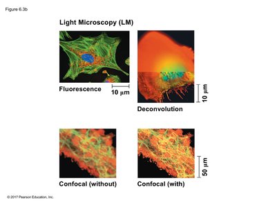
Advanced Light Microscopy Techniques
Fluorescence Microscopy: Uses fluorescent dyes or proteins to label specific cell components, which are then visualized with specific wavelengths of light.
Confocal Microscopy: Uses lasers and optical sectioning to produce sharp images of a single plane within a specimen; can be combined with deconvolution software for enhanced clarity.
Electron Microscopy
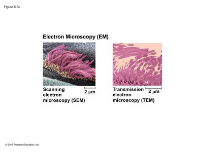
Scanning Electron Microscopy (SEM): Provides detailed 3D images of the surface of a specimen by detecting electrons emitted from the surface.
Transmission Electron Microscopy (TEM): Passes electrons through a thin section of the specimen to reveal internal structures; regions that absorb electrons appear darker.
Both SEM and TEM require non-living specimens due to the preparation process.
Cell Fractionation
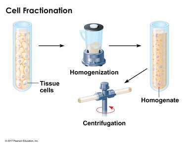
Cell fractionation is a technique used to separate cellular components for detailed study. The process involves homogenizing tissue to release cell contents, then using centrifugation to separate components based on size and density.
Differential Centrifugation
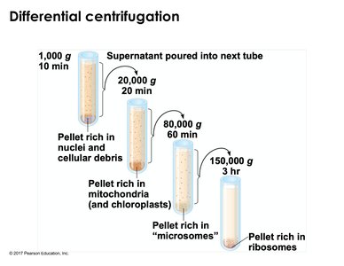
By spinning homogenized cell samples at increasing speeds and durations, different cellular components can be isolated in pellets:
Low speed: nuclei and cellular debris
Medium speed: mitochondria and chloroplasts
High speed: microsomes (fragments of ER, plasma membrane)
Very high speed: ribosomes
Prokaryotic and Eukaryotic Cells
Prokaryotic Cells
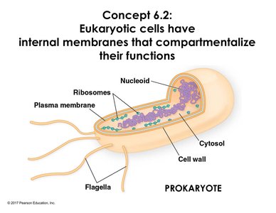
Prokaryotic cells lack a membrane-bound nucleus. Their DNA is located in a region called the nucleoid. They possess a plasma membrane, cytosol, ribosomes, and sometimes flagella for movement. Prokaryotes generally lack extensive internal compartmentalization.
Eukaryotic Cells
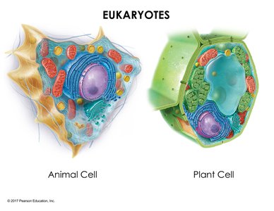
Eukaryotic cells are characterized by internal membranes that compartmentalize functions into organelles. Their DNA is organized into multiple linear chromosomes within a nucleus. Eukaryotic cells are generally larger and more structurally complex than prokaryotic cells.
Plasma Membrane Structure
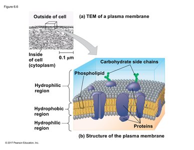
The plasma membrane is a selective barrier composed of a phospholipid bilayer with embedded proteins and carbohydrates. It regulates the passage of substances into and out of the cell and is essential for maintaining cellular homeostasis.
Surface Area to Volume Ratio
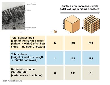
Cell size is limited by the surface area-to-volume ratio. As a cell grows, its volume increases faster than its surface area, reducing the efficiency of material exchange. Cells may have adaptations such as microvilli to increase surface area for absorption.
Small Cube | Large Cube | Many Small Cubes | |
|---|---|---|---|
Total Surface Area | 6 | 150 | 750 |
Total Volume | 1 | 125 | 125 |
Surface-to-Volume Ratio | 6 | 1.2 | 6 |
The Nucleus and Ribosomes
Nucleus
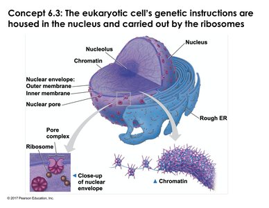
The nucleus is the most prominent organelle in eukaryotic cells, surrounded by a double membrane called the nuclear envelope. Nuclear pores regulate the movement of molecules in and out. The nucleolus within the nucleus is the site of ribosomal RNA synthesis and ribosome assembly.
Ribosomes
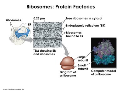
Ribosomes are macromolecular complexes of RNA and protein that synthesize polypeptides. They exist as free ribosomes in the cytosol or bound to the endoplasmic reticulum (ER). Free ribosomes produce proteins for the cytosol, while bound ribosomes synthesize proteins for membranes or secretion. Ribosomal subunits associate and dissociate via non-covalent interactions, allowing flexibility in protein synthesis.
The Endomembrane System
Overview of the Endomembrane System
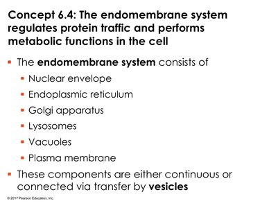
The endomembrane system is a network of membranes within eukaryotic cells that regulates protein traffic and performs metabolic functions. It includes the nuclear envelope, endoplasmic reticulum (ER), Golgi apparatus, lysosomes, vacuoles, and the plasma membrane. Components are connected directly or via vesicle transport.
Endoplasmic Reticulum (ER)
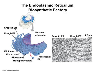
Smooth ER: Lacks ribosomes; involved in lipid synthesis, detoxification, and calcium storage.
Rough ER: Studded with ribosomes; synthesizes proteins for membranes, organelles, or secretion. The interior space is called the lumen, where protein folding and modification (e.g., glycosylation) occur.
Golgi Apparatus
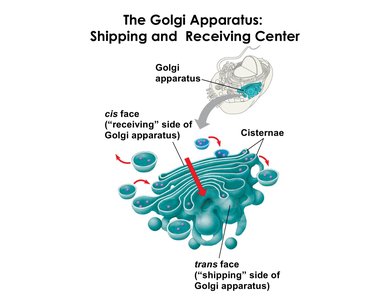
The Golgi apparatus consists of flattened sacs (cisternae) with a cis face (receiving side) and a trans face (shipping side). It modifies, sorts, and packages proteins and lipids for delivery to various destinations.
Lysosomes
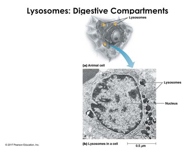
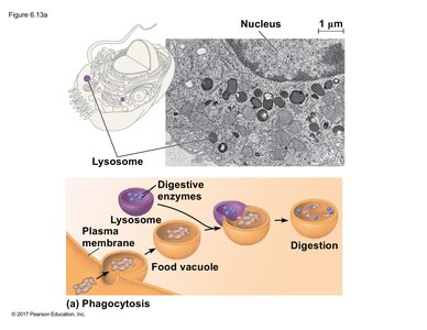
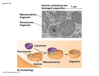
Lysosomes are membrane-bound sacs containing hydrolytic enzymes (acid hydrolases) that digest macromolecules. They function optimally in acidic conditions maintained by proton pumps. Lysosomes are involved in intracellular digestion and recycling of cellular components.
Phagocytosis and Autophagy
Phagocytosis: Cells engulf large particles or other cells, forming a food vacuole (phagosome) that fuses with a lysosome for digestion.
Autophagy: Damaged organelles or cytoplasmic components are enclosed in a membrane (autophagosome) and delivered to lysosomes for degradation and recycling.
Vacuoles
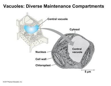
Vacuoles are large membrane-bound organelles prominent in plant cells. They serve as storage sites for water, ions, pigments, proteins, or toxins. In plants, the central vacuole also helps maintain cell rigidity and replaces the lysosomal function found in animal cells.
Integration of the Endomembrane System
The endomembrane system coordinates the synthesis, modification, sorting, and transport of proteins and lipids. Vesicles shuttle materials between organelles, ensuring compartmentalization and efficiency of cellular processes.
