 Back
BackAnimal Responses to the Environment: Nervous System Structure and Function
Study Guide - Smart Notes
 Tailored notes based on your materials, expanded with key definitions, examples, and context.
Animal Responses to the Environment
Introduction
Animals must detect, interpret, and respond to environmental stimuli to survive. These responses are coordinated by the nervous system, which integrates sensory input and generates appropriate motor outputs. Adaptations for environmental response are seen across the animal kingdom, from simple reflexes to complex behaviors.
Exchange and Transport in Circulatory Systems
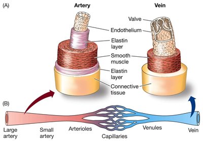
Capillaries and Blood Flow
Capillaries are the primary sites of exchange between blood and tissues. Their structure is specialized to maximize efficient exchange of gases, nutrients, and wastes.
Capillary Walls: Extremely thin (one cell layer) to minimize diffusion distance.
Blood Flow: Slow in capillaries, allowing more time for exchange processes.
Surface Area: Extensive network increases total area for exchange.
Arteries and veins differ structurally and functionally from capillaries. Arteries transport blood away from the heart under high pressure, while veins return blood to the heart and often contain valves to prevent backflow.
Neural Basis of Animal Responses
Types of Neurons
Neurons are specialized cells that transmit information throughout the body using electrical and chemical signals. They are the fundamental units of the nervous system.
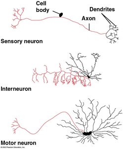
Sensory Neurons: Detect external or internal stimuli (e.g., light, touch, chemical signals) and transmit information to the central nervous system (CNS).
Interneurons: Integrate sensory input and coordinate appropriate responses, primarily located in the CNS.
Motor Neurons: Transmit signals from the CNS to effectors such as muscles or glands, resulting in a response.
Organization of the Nervous System
The nervous system is organized into two main divisions:
Central Nervous System (CNS): Consists of the brain and spinal cord; responsible for processing and integrating information.
Peripheral Nervous System (PNS): Composed of cranial, spinal, and peripheral nerves; transmits sensory and motor information between the CNS and the rest of the body.
Within the PNS:
Afferent (Sensory) Neurons: Carry information to the CNS.
Efferent (Motor) Neurons: Carry information away from the CNS to effectors.
Motor System: Controls voluntary skeletal muscle movements.
Autonomic Nervous System: Regulates involuntary functions (e.g., heartbeat, digestion) and is divided into sympathetic ("fight-or-flight"), parasympathetic ("rest and digest"), and enteric (digestive regulation) branches.
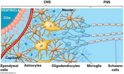
Structure of Neurons and Glial Cells
Neurons have specialized structures for receiving and transmitting signals:
Dendrites: Receive incoming signals from other neurons.
Axon: Conducts electrical impulses away from the cell body.
Synapse: Junction where chemical signals are transmitted to another cell.
Glial cells support, nourish, and protect neurons. Types include astrocytes, oligodendrocytes, microglia, Schwann cells, and ependymal cells.
Diversity in Neuron Structure
Neurons vary greatly in shape and size, reflecting their specialized functions. For example, sensory neurons have long dendrites to receive signals from distant receptors, interneurons have complex branching for integration, and motor neurons have long axons to reach muscles.
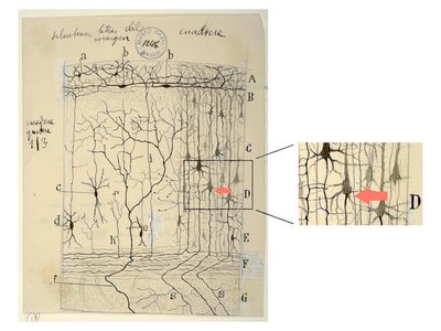
Historical Perspective: Santiago Ramón y Cajal
Santiago Ramón y Cajal was a pioneering neuroscientist who used detailed drawings to reveal the structure of neurons and their connections, laying the foundation for modern neuroscience.
How Neurons Work: Resting Potential and Action Potentials
Electrochemical Gradients and Membrane Potential
Neurons maintain an electrochemical gradient across their membranes, which is essential for generating electrical signals.
Concentration Gradient: Difference in ion concentrations across the membrane.
Electrical Gradient: Difference in charge across the membrane.
Membrane Potential: The voltage difference across the membrane, typically -60 to -80 mV in resting neurons (inside negative relative to outside).
This potential energy is used to transmit signals rapidly along the neuron.
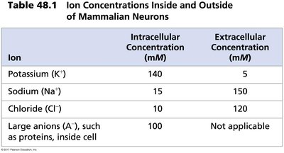
Ion Concentrations in Neurons
The distribution of ions inside and outside the neuron is maintained by selective ion channels and active transport (Na+/K+ pump).
Ion | Intracellular Concentration (mM) | Extracellular Concentration (mM) |
|---|---|---|
Potassium (K+) | 140 | 5 |
Sodium (Na+) | 15 | 150 |
Chloride (Cl-) | 10 | 120 |
Large anions (A-), e.g., proteins | 100 | Not applicable |
Gated Ion Channels and Action Potentials
Neurons contain gated ion channels that open or close in response to stimuli, altering the membrane potential and generating action potentials.
Resting Potential: Maintained by Na+/K+ pumps and selective permeability of the membrane.
Depolarization: Inside of the cell becomes less negative (more positive) due to Na+ influx.
Hyperpolarization: Inside of the cell becomes more negative, often due to K+ efflux.
Action Potential: A rapid, self-propagating change in membrane potential that travels along the axon. It is an all-or-none response, triggered only if the threshold is reached (about -55 mV).
Steps of an Action Potential:
Depolarization: Na+ channels open, Na+ enters the cell.
Repolarization/Hyperpolarization: K+ channels open, K+ leaves the cell.
Undershoot: Membrane potential temporarily becomes more negative than resting potential.
Restoration: Na+/K+ pump restores resting potential.
Equation for Nernst Potential (for a single ion):
Where: R = gas constant, T = temperature (K), z = charge of ion, F = Faraday's constant.
Summary
Animal responses to the environment are coordinated by the nervous system, which relies on specialized cells (neurons and glia), organized structures (CNS and PNS), and electrochemical gradients to detect, integrate, and respond to stimuli. Understanding these processes is fundamental to biology and medicine.
