 Back
BackEukaryotic and Prokaryotic Cells, Membrane Transport, and Diffusion/Osmosis
Study Guide - Smart Notes
 Tailored notes based on your materials, expanded with key definitions, examples, and context.
Topic 4: Eukaryotic and Prokaryotic Cells
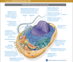
Eukaryotic Cells
Eukaryotic cells are complex cells characterized by the presence of membrane-bound organelles and a defined nucleus. These cells are found in animals, plants, fungi, and protists.
Membrane-bound organelles: Specialized structures such as the Golgi body, endoplasmic reticulum, and mitochondria perform distinct cellular functions.
Membrane-bound nucleus: Contains the cell's genetic material (DNA) organized into linear chromosomes.
Other features: Eukaryotic cells have a plasma membrane, cytoskeleton, and ribosomes.
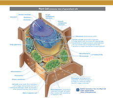
Plant Cells
Plant cells are a type of eukaryotic cell with unique structures that support their function in photosynthesis and storage.
Large central vacuole: Stores water, pigments, and waste products.
Plastids: Organelles such as chloroplasts (for photosynthesis) and chromoplasts (for pigment storage).
Cell wall: Rigid structure that surrounds and protects the plasma membrane.
Lack centrioles and lysosomes: Unlike animal cells, plant cells do not have these organelles.
Comparison: Eukaryotes vs. Prokaryotes, Animal vs. Plant Cells
The following table summarizes the key differences among eukaryotes, prokaryotes, animal cells, and plant cells:
Feature | Eukaryotes | Prokaryotes | Animal Cells | Plant Cells |
|---|---|---|---|---|
Nucleus | Present | Absent (nucleoid region) | Present | Present |
Membrane-bound organelles | Present | Absent | Present | Present |
Chromosomes | Linear | Circular | Linear | Linear |
Cell wall | Some (plants, fungi) | Present | Absent | Present |
Central vacuole | Some (plants) | Absent | Absent | Present |
Centrioles | Some (animals) | Absent | Present | Absent |
Lysosomes | Some (animals) | Absent | Present | Absent |
Cytochemical Staining
Cytochemical staining is used to visualize specific cell components under the microscope, which are otherwise difficult to see.
Targeted staining: Stains can highlight DNA, RNA, glycogen, lipids, etc.
Enzyme treatment: Enzymes such as DNase, RNase, amylase, and lipase can remove specific components to confirm the stain's specificity.
Example: Amylase removes glycogen; observing before and after staining confirms glycogen presence.
Microscopy and Slide Preparation
Microscopy is essential for studying cell structure. Different microscopes are used for different purposes:
Compound light microscope: Used for viewing thin sections of cells and tissues at high magnification.
Dissecting microscope: Used for viewing larger, three-dimensional specimens at lower magnification.
Electron microscope: Provides high-resolution images of cell ultrastructure.
Proper slide preparation and staining are crucial for clear observation. For example, Lugol’s iodine stains starch in plant cells, and methylene blue stains nuclei in animal cells.
Topic 5: Diffusion, Osmosis, and Membrane Transport
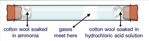
Diffusion
Diffusion is the passive movement of molecules from an area of high concentration to an area of low concentration, driven by the concentration gradient.
Passive process: Does not require energy input.
Factors affecting rate: Diffusion is faster in gases than in liquids, and in liquids than in solids. Higher temperatures and smaller molecules increase the rate of diffusion.
Example: Ammonia (NH3) diffuses faster than hydrochloric acid (HCl) due to its lower molecular weight.
Osmosis
Osmosis is the diffusion of water across a selectively permeable membrane from an area of higher water potential to lower water potential.
Semi-permeable membrane: Allows water to pass but restricts solute movement.
Solvent: In biological systems, water is the solvent.
Solute: Substances dissolved in water, such as salts and sugars.
Water and Chemical Potential
Chemical potential is the free energy available to move molecules. Water potential is the potential energy of water compared to pure water at constant temperature and pressure.
Pure water: Maximum water potential (zero).
Solutes: Adding solutes makes water potential more negative, restricting water movement.
Movement: Water moves from regions of higher (less negative) to lower (more negative) water potential.
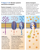
Membrane Transport Mechanisms
Cells use different mechanisms to transport substances across membranes:
Passive transport: Includes diffusion and osmosis; does not require energy. Molecules move down their concentration gradient.
Active transport: Requires energy (ATP) to move molecules against their concentration gradient.
Dialysis Tubing Experiment
Dialysis tubing is a model for a semi-permeable membrane, allowing small molecules like glucose to pass but blocking larger molecules like starch.
Starch test: Lugol’s iodine turns blue-black in the presence of starch.
Glucose test: Benedict’s reagent changes color when glucose is present.
Results: Glucose diffuses through the tubing; starch does not. Over time, glucose concentrations equilibrate inside and outside the tubing.
Summary Table: Membrane Types
Membrane Type | Example | Pore Properties | Function |
|---|---|---|---|
Selectively permeable | Cell membrane | Dynamic, can change pore size | Regulates entry/exit of substances |
Semi-permeable | Dialysis tubing | Fixed pore size | Allows certain molecules to pass |
Key Equations
Water potential: Where is total water potential, is solute potential, and is pressure potential.
Microscopy Best Practices
Always start with the lowest magnification (4x) to locate your specimen.
Use oil only with the 100x objective and clean with lens paper and appropriate cleaner after use.
Proper cleaning and handling of microscopes are essential for clear observations and instrument longevity.
