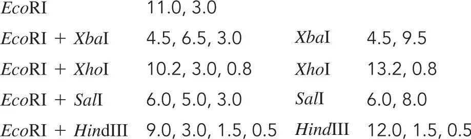
You have recovered a cloned DNA segment from a vector and determine that the insert is 1300 bp in length. To characterize this cloned segment, you isolate the insert and decide to construct a restriction map. Using enzyme I and enzyme II, followed by gel electrophoresis, you determine the number and size of the fragments produced by enzymes I and II alone and in combination, as recorded in the following table. Construct a restriction map from these data, showing the positions of the restriction-enzyme cutting sites relative to one another and the distance between them in units of base pairs.
Table of contents
- 1. Introduction to Genetics51m
- 2. Mendel's Laws of Inheritance3h 37m
- 3. Extensions to Mendelian Inheritance2h 41m
- 4. Genetic Mapping and Linkage2h 28m
- 5. Genetics of Bacteria and Viruses1h 21m
- 6. Chromosomal Variation1h 48m
- 7. DNA and Chromosome Structure56m
- 8. DNA Replication1h 10m
- 9. Mitosis and Meiosis1h 34m
- 10. Transcription1h 0m
- 11. Translation58m
- 12. Gene Regulation in Prokaryotes1h 19m
- 13. Gene Regulation in Eukaryotes44m
- 14. Genetic Control of Development44m
- 15. Genomes and Genomics1h 50m
- 16. Transposable Elements47m
- 17. Mutation, Repair, and Recombination1h 6m
- 18. Molecular Genetic Tools19m
- 19. Cancer Genetics29m
- 20. Quantitative Genetics1h 26m
- 21. Population Genetics50m
- 22. Evolutionary Genetics29m
18. Molecular Genetic Tools
Genetic Cloning
Problem 18
Textbook Question
To further analyze the CRABS CLAW gene, you create a map of the genomic clone. The 11-kb EcoRI fragment is ligated into the EcoRI site of the MCS of the vector shown in Problem 18. You digest the double-stranded form of the genome with several restriction enzymes and obtain the following results. Draw, as far as possible, a map of the genomic clone of CRABS CLAW.

What restriction digest would help resolve any ambiguity in the map?

1
Step 1: Begin by analyzing the data provided for each restriction enzyme digest. Note the fragment sizes generated by EcoRI, XbaI, XhoI, SalI, and HindIII individually, as well as the combinations of EcoRI with each enzyme. This will help identify overlapping fragments and potential cut sites.
Step 2: Construct a preliminary map of the genomic clone based on the fragment sizes. Start with the EcoRI digest, which produces fragments of 11.0 kb and 3.0 kb. Place these fragments on the map, ensuring their total length matches the genomic clone size.
Step 3: Incorporate the results of the double digests (e.g., EcoRI + XbaI, EcoRI + XhoI, etc.) into the map. For example, the EcoRI + XbaI digest produces fragments of 4.5 kb, 6.5 kb, and 3.0 kb. Use this information to determine the relative positions of XbaI cut sites within the EcoRI fragments.
Step 4: Repeat the process for the other double digests (EcoRI + XhoI, EcoRI + SalI, EcoRI + HindIII). Compare the fragment sizes generated by each combination to refine the map and resolve ambiguities in the placement of restriction sites.
Step 5: To resolve any remaining ambiguities, consider performing additional restriction digests with enzymes not yet used or combinations of enzymes that target specific regions of the map. For example, a digest with XbaI + XhoI or SalI + HindIII could provide further clarity on overlapping fragments or closely spaced cut sites.

This video solution was recommended by our tutors as helpful for the problem above
Video duration:
2mPlay a video:
Was this helpful?
Key Concepts
Here are the essential concepts you must grasp in order to answer the question correctly.
Restriction Enzymes
Restriction enzymes, or restriction endonucleases, are proteins that cut DNA at specific sequences, allowing scientists to manipulate and analyze genetic material. Each enzyme recognizes a unique sequence of nucleotides, which is crucial for creating DNA fragments of desired lengths for cloning or mapping. Understanding how these enzymes work is essential for interpreting the results of a restriction digest and constructing a genomic map.
Recommended video:
Guided course
Mapping with Markers
Genomic Mapping
Genomic mapping involves determining the locations of genes and other important sequences on a chromosome. This process often utilizes restriction enzyme digests to create a physical map of DNA fragments, which can be analyzed to understand gene organization and function. A well-constructed map helps resolve ambiguities in gene locations and relationships, making it a fundamental aspect of genetic analysis.
Recommended video:
Guided course
Modern Mapping
Fragment Size Analysis
Fragment size analysis is a technique used to determine the lengths of DNA fragments generated by restriction enzyme digestion. By comparing the sizes of these fragments, researchers can infer the positions of restriction sites and the overall structure of the genomic clone. This analysis is critical for resolving ambiguities in genomic maps, as different combinations of restriction enzymes can yield distinct patterns that clarify the arrangement of genetic elements.
Recommended video:
Guided course
Chi Square Analysis
Related Videos
Related Practice
Textbook Question
659
views