 Back
BackBlood, Lymphatic, and Immune Systems: Medical Terminology Essentials
Study Guide - Smart Notes
 Tailored notes based on your materials, expanded with key definitions, examples, and context.
The Blood, Lymphatic, and Immune Systems
Introduction
This chapter explores the essential terminology, structure, and function of the blood, lymphatic, and immune systems. Understanding these systems is crucial for comprehending how the body maintains homeostasis, defends against disease, and transports vital substances.
Blood: Components and Functions
Definition and General Properties
Blood is a specialized connective tissue circulating in a closed system (heart and blood vessels).
It is unique among connective tissues because it is in liquid form.
Color: Arterial blood is bright red; venous blood is dark red.
pH: Slightly alkaline (7.35–7.45).
Viscosity: High.
Temperature: 38ºC (100.4ºF).
Volume: Adult male: 5–6 L; Adult female: 4–5 L.
Functions of Blood
Maintains homeostasis.
Transports nutrients, vitamins, minerals, waste products, hormones, and gases.
Protects against foreign substances (microorganisms, toxins).
Forms clots (coagulation) to prevent blood loss.
Composition of Blood
Plasma (about 55%): Fluid portion containing water (92%), plasma proteins (7%), and other solutes (1%).
Formed Elements (about 45%): Cellular components including red blood cells (RBCs), white blood cells (WBCs), and platelets.
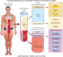
Plasma Proteins
Albumins: Most abundant; regulate colloidal osmotic pressure and transport substances.
Globulins: Include antibodies (immunoglobulins) for immunity.
Fibrinogen: Essential for blood clotting; forms fibrin threads in clots.
Formed Elements: Blood Cells
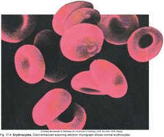
Red Blood Cells (Erythrocytes)
Transport oxygen and carbon dioxide.
Biconcave shape increases surface area for gas exchange.
Mature RBCs lack a nucleus and most organelles; filled with hemoglobin (Hb).
Life span: ~120 days.
Normal values: Men: 13.5–17.5 g/dL Hb; Women: 12.0–15.5 g/dL Hb.
Hemoglobin
Iron-containing protein responsible for oxygen transport and red color of blood.
Abbreviated as Hgb or Hb.
Low hemoglobin levels indicate anemia.
Disorders of RBCs
Anemia: Reduced oxygen-carrying capacity of blood.
Iron Deficiency Anemia: Most common; due to low iron.
Pernicious Anemia: Vitamin B12 deficiency (often autoimmune).
Sickle Cell Anemia: Genetic; abnormal, rigid RBC shape.
Aplastic Anemia: Bone marrow fails to produce RBCs.
Hemolytic Anemia: RBCs destroyed faster than produced.
Thalassemia: Reduced hemoglobin production.
Polycythemia: Overproduction of RBCs by bone marrow.
White Blood Cells (Leukocytes)
Part of the immune system; defend against infection and disease.
Normal range: 4,000–11,000 cells/µL.
Two main groups:
Granulocytes: Neutrophils, Eosinophils, Basophils.
Agranulocytes: Lymphocytes, Monocytes.
Granulocytes
Neutrophils: Most abundant; first responders; phagocytosis of bacteria.
Eosinophils: Fight parasites; involved in allergic reactions.
Basophils: Release histamine (inflammation/allergy) and heparin (anticoagulant).
Agranulocytes
Lymphocytes: B cells (produce antibodies), T cells (attack infected cells); adaptive immunity.
Monocytes: Largest WBCs; become macrophages in tissues; phagocytosis of pathogens and debris.
Disorders of WBCs
Leukocytosis: Excessive WBC count.
Leukemia: Cancer with high WBC count.
Leukopenia: Low WBC count.
Infectious Mononucleosis: Caused by Epstein-Barr virus; common in ages 15–25.
Platelets (Thrombocytes)
Small cell fragments essential for blood clotting (hemostasis).
Produced in bone marrow from megakaryocytes.
Thrombocytopenia: Low platelets; risk of bleeding.
Thrombocytosis: High platelets; risk of clots.
Disorders of Coagulation
Hemophilia: Deficiency of clotting factors (e.g., Factor VIII).
Thrombus: Clot attached to vessel wall.
Embolus: Movable clot.
Purpura: Purple/red skin spots from bleeding under the skin.
Bruises: Blood leakage from vessels.
Hemostasis (Stopping Bleeding)
Vascular spasm: Vasoconstriction reduces blood flow after injury.
Platelet plug formation: Platelets adhere to damaged area.
Blood coagulation: Clotting factors convert fibrinogen to fibrin, stabilizing the plug.
Clot retraction and repair: Platelets contract to shrink the clot.
Fibrinolysis: Plasmin dissolves the clot after healing.
Blood Groups
ABO System: Based on presence of A and B antigens on RBCs.
Rh Factor: Rh⁺ (D antigen present), Rh⁻ (D antigen absent).
Erythroblastosis fetalis: Hemolytic disease of newborn due to Rh incompatibility.
Antigens and Antibodies
Antigen: Molecule on RBC surface (e.g., ABO, Rh).
Antibody: Protein in plasma that binds specific antigen.
Agglutination: Clumping due to antigen-antibody reaction (e.g., mismatched transfusion).
Diagnostic Procedures
CBC: Complete blood count.
DIFF: Differential WBC count.
PT: Prothrombin time (clotting test).
Hematocrit: Percentage of RBC volume.
Lymphatic System
Functions
Absorbs excess interstitial fluid and returns it to the bloodstream.
Removes foreign chemicals, cells, and debris from tissues.
Absorbs dietary lipids from the small intestine.
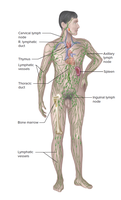
Components
Lymphatic vessels (lymphatics): Transport lymph.
Lymph: Clear, colorless fluid.
Lymph nodes: Filter lymph; contain immune cells.
Lymphatic ducts and trunks: Large vessels draining lymph into veins.
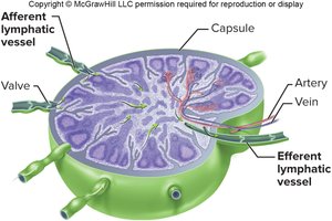
Lymph Nodes
Bean- or kidney-shaped organs enclosed in a fibrous capsule.
Divided into cortex (outer) and medulla (inner), rich in B and T lymphocytes.
Filter lymph, trapping bacteria, viruses, and cancer cells; immune cells destroy these before lymph returns to blood.
Major Lymphatic Organs
Spleen: Filters blood; located in upper left abdomen.
Tonsils: Three types (palatine, pharyngeal/adenoid, lingual); protect throat; tonsillectomy is surgical removal.
Thymus: Upper chest; essential for T-lymphocyte development; secretes thymosin; large in children, shrinks after puberty.
Flow of Lymph
Pathway: Interstitial fluid → lymphatic capillaries → lymphatic vessels → lymph nodes → lymphatic trunks → right lymphatic duct/thoracic duct → subclavian veins.
Thoracic duct: Drains most of the body into left subclavian vein.
Right lymphatic duct: Drains right upper body into right subclavian vein.
Immune System
Immunity
Immunity: Ability to resist harmful effects of microorganisms and foreign substances.
Innate Immunity: Inborn, non-specific; includes skin, mucous membranes, phagocytes, NK cells, inflammation, fever.
Adaptive Immunity: Specific; develops after exposure to antigens.
Cellular (cell-mediated) immunity: T lymphocytes attack infected cells.
Humoral (antibody-mediated) immunity: B lymphocytes produce antibodies.
Disorders of the Immune System
Immunodeficiency: Example: AIDS (caused by HIV).
Autoimmune Disorders: Immune system attacks own tissues (e.g., rheumatoid arthritis, lupus, type 1 diabetes).
Hypersensitivity (Allergic) Reactions: Overactive immune response (e.g., anaphylaxis, hay fever, asthma).
