 Back
BackOncology: Medical Terminology and Cancer Fundamentals
Study Guide - Smart Notes
 Tailored notes based on your materials, expanded with key definitions, examples, and context.
Oncology and the Cellular Basis of Cancer
Introduction to Oncology
Oncology is the medical specialty focused on the study, diagnosis, and treatment of cancer. Unlike other specialties, oncology is not limited to a single body system, as cancer can arise in any tissue or organ. Understanding cancer requires knowledge of normal cell structure, cell division, and the mechanisms by which cells become cancerous.
Cell Structure and Function
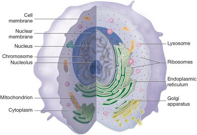
Structures of a Normal Cell
Cells are the smallest independently functioning units of the body, capable of division and essential for life. Each cell contains specialized structures (organelles) that perform vital functions such as energy production, protein synthesis, and defense against pathogens.
Cell membrane: Regulates entry and exit of substances.
Nucleus: Contains genetic material (chromosomes) and controls cellular activities.
Chromosomes: Structures composed of DNA; humans have 46 chromosomes (23 pairs).
Nucleolus: Site of ribosome synthesis within the nucleus.
Mitochondrion: Produces cellular energy (ATP).
Cytoplasm: Gel-like substance containing organelles.
Lysosome: Digests cellular waste.
Ribosomes: Synthesize proteins.
Endoplasmic reticulum: Synthesizes and transports proteins and lipids.
Golgi apparatus: Modifies, sorts, and packages proteins.
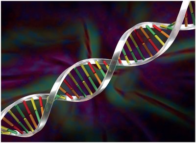
Genetic Material: DNA and Chromosomes
DNA (deoxyribonucleic acid) is the hereditary material in cells, organized into chromosomes. Each chromosome contains one long DNA molecule, which is divided into genes. Genes encode the instructions for making proteins, which determine cell structure and function.
Gene: A segment of DNA that codes for a specific protein.
DNA double helix: The structure of DNA, consisting of two strands wound around each other, with paired bases forming the rungs.
Cell Division and Cancer Development
Normal Cell Division
Normal cells divide by mitosis, a controlled process that ensures tissue growth and repair. Cell division is regulated by growth factors and suppressor genes, which prevent excessive proliferation. Meiosis is a specialized form of cell division that produces gametes (sperm and egg cells) with half the normal chromosome number.
Mitosis: Produces two identical daughter cells for growth and repair.
Meiosis: Produces gametes with 23 chromosomes each, ensuring genetic diversity.
Suppressor genes: Inhibit excessive cell division.
Cancer Cell Division and Growth
Cancer arises when normal regulatory mechanisms fail, leading to uncontrolled cell division. Cancer cells often lose their specialized functions (differentiation) and revert to an immature, undifferentiated state (anaplasia). Abnormal cell growth can be classified as:
Hyperplasia: Increased number of normal cells.
Dysplasia: Abnormal cell size, shape, and arrangement.
Anaplasia: Complete loss of differentiation; cells appear embryonic.
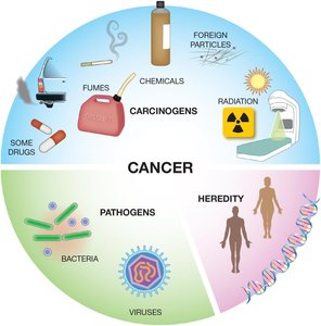
Causes of Cancer
Cancer can be caused by a variety of factors, including environmental carcinogens, pathogens, and hereditary mutations. Carcinogens are substances that induce genetic mutations, while certain viruses and bacteria can also trigger cancer development. Inherited genetic mutations (e.g., in tumor suppressor genes like p53) significantly increase cancer risk.
Carcinogens: Chemicals, radiation, fumes, and some drugs.
Pathogens: Bacteria and viruses.
Heredity: Inherited gene mutations.
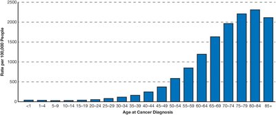
Cancer Incidence Across the Lifespan
The risk of developing cancer increases with age. While cancer is rare in children and young adults, incidence rises sharply in older populations, with nearly 40% of people diagnosed at some point in their lives.
Medical Terminology in Oncology
Key Terms and Definitions
Anaplasia: Condition where mature, differentiated cells become undifferentiated and cancerous.
Carcinoid tumor: Slow-growing tumor, often in the digestive tract, rarely metastasizes.
Carcinomatosis: Presence of cancerous tumors at multiple sites in the body.
Dysplasia: Abnormal cell growth, not yet cancerous.
Lymphadenopathy: Enlarged lymph nodes, often due to cancer spread.
Precancerous: Cells/tissues with abnormal features, not yet cancerous.
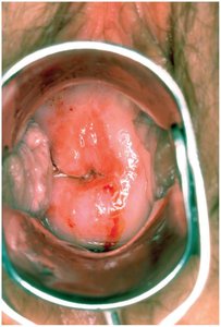
Cervical Dysplasia Example
Cervical dysplasia is a condition where abnormal cells are found on the cervix, which may develop into cancer if untreated. It is often detected during routine gynecological exams.
Characteristics of Cancer Cells and Tumors
Eight Key Characteristics
Cancer cells and tumors exhibit several distinguishing features:
Do not contribute to normal body function.
Are undifferentiated and lack specialized functions.
Are disorganized in arrangement.
Divide more rapidly than normal cells.
Form irregular, unencapsulated solid tumors.
Induce angiogenesis (growth of new blood vessels).
Are invasive, infiltrating surrounding tissues.
Can metastasize (spread) to distant sites via blood or lymph.
Metastasis
Metastasis is the process by which cancer cells break away from the primary tumor and travel through the bloodstream or lymphatic system to form new tumors in other organs.
Types of Cancer and Body Systems
Classification by Tissue or Organ
Cancers are classified based on the tissue or organ of origin. Common types include:
Carcinoma: Cancer of epithelial cells (skin, mucous membranes).
Sarcoma: Cancer of connective tissues (muscle, bone, fat).
Leukemia: Cancer of blood-forming tissues (white blood cells).
Lymphoma: Cancer of lymphatic tissue.
Myeloma: Cancer of plasma cells in bone marrow.
Special Cancer Types
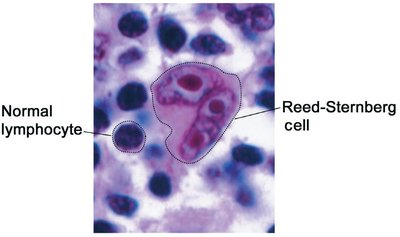
Reed-Sternberg cell: Atypical cell found in Hodgkin lymphoma, diagnostic for this disease.
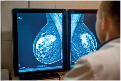
Adenocarcinoma of the Breast
Adenocarcinoma is a cancer of glandular epithelial cells, such as those in the breast. It often appears as a dense, irregular mass on mammography.
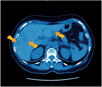
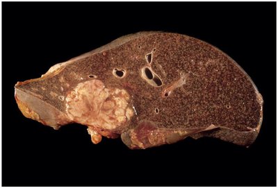
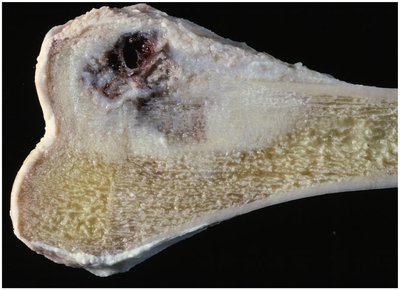
Liver Cancer Example
Liver cancer can originate in the ducts or hepatocytes and often presents as a large, irregular tumor invading normal tissue.
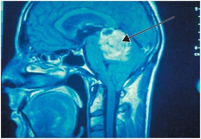
Astrocytoma (Glioma)
Astrocytoma is a type of glioma, a cancer of the brain's supporting cells (astrocytes). Glioblastoma multiforme is a highly malignant form.
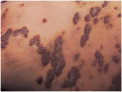
Kaposi Sarcoma
Kaposi sarcoma is a cancer of the skin and subcutaneous tissue, often seen in immunocompromised patients (e.g., AIDS).
Osteosarcoma
Osteosarcoma is a malignant tumor of bone, commonly affecting children and adolescents. It often invades surrounding soft tissues.
Diagnostic and Laboratory Procedures
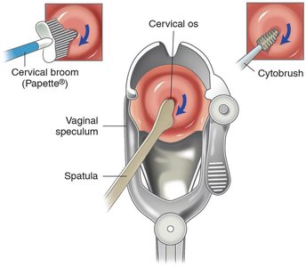
Cytology and Screening Tests
Cytology tests examine cells for abnormalities. The Pap test is a common screening tool for cervical cancer, involving the collection of cervical cells for microscopic examination.
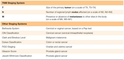
Cancer Staging Systems
Cancer staging describes the extent of disease spread. The TNM system is widely used:
T (Tumor): Size of the primary tumor.
N (Nodes): Number of regional lymph nodes involved.
M (Metastasis): Presence of distant metastases.
Other staging systems exist for specific cancers (e.g., FIGO for gynecologic cancers, Gleason score for prostate cancer).
System | Purpose |
|---|---|
TNM | Primary tumor size, lymph node involvement, metastasis |
Bethesda | Cervical/vaginal cancer (Pap test) |
CIN | Cervical intraepithelial neoplasia |
Clark/Breslow | Malignant melanoma |
Dukes | Colon/rectal cancer |
FIGO | Ovarian/uterine cancer |
Gleason | Prostate cancer |
Jewett-Whitmore | Prostate cancer |
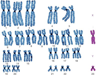
Karyotype Analysis
Karyotyping examines the number and structure of chromosomes, identifying genetic abnormalities associated with cancer.
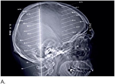
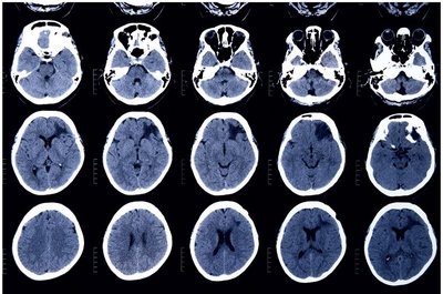
Radiology and Imaging
Imaging techniques such as CT (computed tomography) and MRI (magnetic resonance imaging) are essential for locating tumors and assessing metastasis.
CT scan: Uses X-rays to create cross-sectional images ('slices') of the body.
MRI: Uses magnetic fields and radio waves for detailed images without radiation exposure.
Cancer Treatment Modalities
Chemotherapy
Chemotherapy uses drugs to target rapidly dividing cancer cells. Side effects include hair loss, as hair follicle cells also divide rapidly. Adjuvant therapy refers to chemotherapy given after surgery to eliminate residual cancer cells.
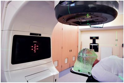
Radiation Therapy
Radiation therapy uses high-energy waves or particles to destroy cancer cells. It can be delivered externally (external beam radiotherapy) or internally (brachytherapy). Cancers vary in their sensitivity to radiation (radiosensitive vs. radioresistant).
Surgical Procedures
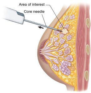
Surgery is often used to diagnose (biopsy) and treat cancer. A core needle biopsy removes tissue samples for analysis, often guided by imaging.
Abbreviations in Oncology
Common Abbreviations
Bx: Biopsy
Ca: Cancer; carcinoma
CT: Computed tomography
CBC: Complete blood count
ER/PR: Estrogen/progesterone receptor
HCG: Human chorionic gonadotropin
MRI: Magnetic resonance imaging
PICC: Peripherally inserted central catheter
PSA: Prostate-specific antigen
TNM: Tumor, nodes, metastases (staging system)
Summary
Understanding oncology requires a foundation in cell biology, genetics, and medical terminology. Cancer is a complex group of diseases with diverse causes, characteristics, and treatments. Mastery of the terminology and concepts outlined above is essential for students preparing for exams and future clinical practice.
