 Back
BackOrthopedics and the Skeletal System: Medical Terminology and Clinical Concepts
Study Guide - Smart Notes
 Tailored notes based on your materials, expanded with key definitions, examples, and context.
Orthopedics and the Skeletal System
Introduction to Orthopedics
Orthopedics is the medical specialty focused on the anatomy, physiology, and diseases of the skeletal and muscular systems. Orthopedists diagnose and treat conditions using medical, surgical, and pharmacological interventions.
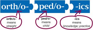
Orthopedics derives from Greek roots: orth/o (straight), ped/o (child), and -ics (knowledge; practice).
Orthopedists address both congenital and acquired disorders of bones and muscles.
Anatomy of the Skeletal System
Overview of the Skeletal System
The human skeletal system consists of 206 bones and associated structures, providing support, protection, and movement for the body.
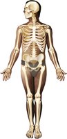
Divided into the axial skeleton (central axis: skull, vertebral column, rib cage) and appendicular skeleton (limbs and girdles).
Functions include support, protection of organs, movement, mineral storage, and blood cell production.
Axial Skeleton
The axial skeleton forms the central structure of the body, including the skull, vertebral column, and thoracic cage.
Skull: Composed of cranial and facial bones.
Vertebral column: Supports the head and trunk, protects the spinal cord.
Thoracic cage: Protects the heart and lungs.
Cranial Bones
The cranium consists of eight bones that protect the brain and form the structure of the head.
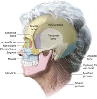
Frontal bone: Forehead
Parietal bones (2): Upper sides and posterior
Occipital bone: Base of the skull
Temporal bones (2): Sides of the skull
Sphenoid bone: Central base
Ethmoid bone: Nasal septum
Facial Bones
Fourteen facial bones support the nose, cheeks, and lips, and form the structure of the face.
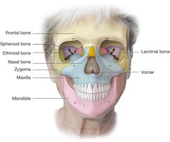
Nasal bones (2): Bridge of the nose
Vomer: Inferior nasal septum
Inferior nasal conchae (2): Lateral walls of nasal cavity
Lacrimal bones (2): Eye sockets near tear glands
Zygomatic bones (2): Cheekbones
Maxilla (2 fused): Upper jaw
Palatine bones (2): Hard palate
Mandible: Lower jaw, only moveable skull bone
Fontanels
Fontanels are soft spots on an infant's skull where cranial bones have not yet fused, allowing for brain growth and flexibility during birth.
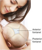
Anterior fontanel: Large soft spot between frontal bones
Posterior fontanel: Smaller soft spot between parietal bones
Ossicles and Hyoid Bone
Ossicles: Three tiny bones (malleus, incus, stapes) in each middle ear, essential for hearing.
Hyoid bone: Located in the neck, not attached to other bones; supports tongue and muscles of the mouth and throat.
Thoracic Cage
The thoracic cage protects vital organs and supports the upper body.
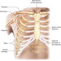
Sternum: Central bone with manubrium, body, and xiphoid process
Ribs: 12 pairs (1-7 true, 8-10 false, 11-12 floating)
Vertebral Column
The vertebral column consists of 33 vertebrae in five regions, providing structural support and protecting the spinal cord.
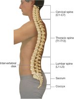
Cervical (C1–C7): Neck region
Thoracic (T1–T12): Upper back
Lumbar (L1–L5): Lower back
Sacrum: Five fused vertebrae
Coccyx: Four fused vertebrae (tailbone)
Appendicular Skeleton
The appendicular skeleton includes the bones of the limbs and girdles, facilitating movement and interaction with the environment.
Shoulder Girdle
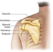
Clavicle: Collarbone, connects sternum to scapula
Scapula: Shoulder blade, articulates with humerus
Upper Extremity
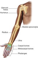
Humerus: Upper arm bone
Radius and Ulna: Forearm bones
Carpals: Eight wrist bones
Metacarpals: Five hand bones
Phalanges: Finger bones (three per finger, two in thumb)
Pelvic Girdle
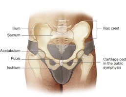
Ilium: Superior hip bone
Ischium: Inferior hip bone
Pubis: Anterior hip bone
All three form the acetabulum (hip socket)
Lower Extremity
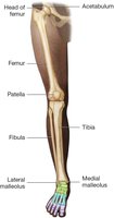
Femur: Thigh bone
Tibia: Shin bone
Fibula: Thin lateral bone of the lower leg
Patella: Kneecap
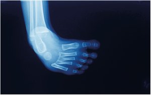
Ankle and Foot
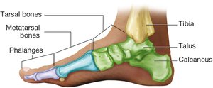
Tarsals: Seven ankle bones
Metatarsals: Five midfoot bones
Phalanges: Toe bones (three per toe, two in hallux/great toe)
Structure of Bone
Bone Tissue and Structure
Bones are composed of osseous tissue, a specialized connective tissue. The structure of a long bone includes several key regions and tissues.
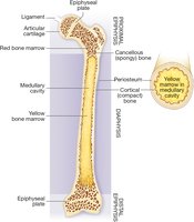
Periosteum: Outer fibrous membrane
Diaphysis: Shaft of the bone
Epiphysis: Ends of the bone
Cortical (compact) bone: Dense outer layer
Cancellous (spongy) bone: Less dense, found at ends
Medullary cavity: Contains yellow bone marrow (fat storage)
Red bone marrow: Site of blood cell production
Joints, Cartilage, and Ligaments
Types of Joints
Joints (articulations) are connections between bones that allow for varying degrees of movement.
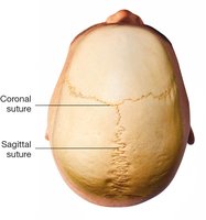
Suture joints: Immovable, found in the skull
Symphysis joints: Slightly moveable, e.g., pubic symphysis, intervertebral discs
Synovial joints: Freely moveable, include hinge (elbow, knee) and ball-and-socket (shoulder, hip)
Joint Structure
Articular cartilage: Covers bone ends in synovial joints
Meniscus: Cartilage pad in some synovial joints
Ligaments: Strong connective tissue bands stabilizing joints
Synovial membrane: Produces lubricating synovial fluid
Physiology of Bone Growth and Remodeling
Bone Cells and Remodeling
Osteocytes: Mature bone cells maintaining mineral content
Osteoclasts: Break down bone tissue, releasing calcium
Osteoblasts: Build new bone matrix
Osteogenesis: Process of new bone formation
Bone remodeling is a continuous process, with about 10% of the skeleton replaced each year. In children, bone formation exceeds breakdown; in adults, the rates are balanced; in older adults, breakdown exceeds formation.
Bone and Cartilage Diseases
Types of Bone Fractures
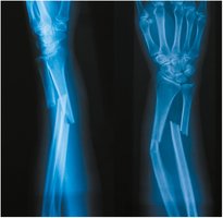
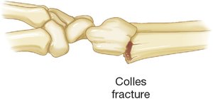
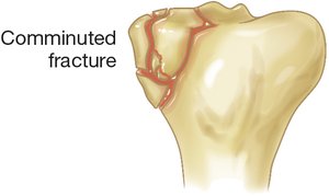
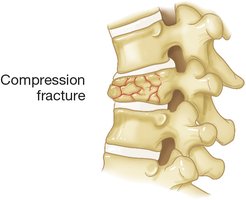
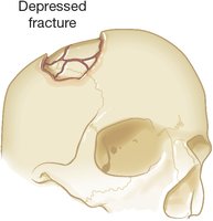
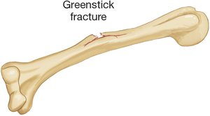
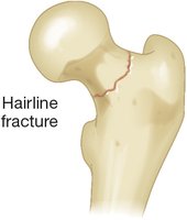
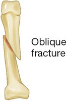
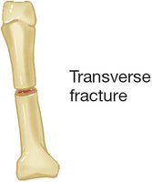
Fractures are breaks in bone integrity, classified by their pattern and severity.
Type of Fracture | Description |
|---|---|
Colles Fracture | Distal radius fracture, often from a fall |
Comminuted Fracture | Bone shattered into multiple pieces |
Compression Fracture | Bone is crushed, common in vertebrae |
Depressed Fracture | Bone pressed inward, typically in skull |
Greenstick Fracture | Bone bends and cracks, common in children |
Hairline Fracture | Thin fracture, bone segments remain aligned |
Oblique Fracture | Diagonal break across the bone |
Spiral Fracture | Twisting force causes spiral break |
Transverse Fracture | Break is perpendicular to bone's axis |
Osteoporosis
Osteoporosis is a disease characterized by decreased bone density and increased fracture risk.
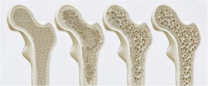
Bone becomes porous and fragile due to loss of mineral content.
Common in postmenopausal women and elderly individuals.
Chest and Spine Diseases
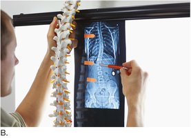
Scoliosis
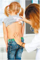
Scoliosis is a lateral curvature of the spine, often diagnosed in childhood or adolescence.
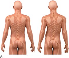
May cause uneven shoulders, hips, and arm lengths.
Diagnosis involves physical examination and imaging.
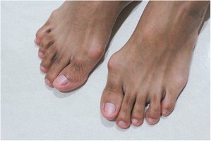
Hallux Valgus and Bunion
Hallux valgus is a lateral deviation of the great toe, often associated with a bunion (painful swelling at the base of the toe).
Clubfoot
Clubfoot (talipes equinovarus) is a congenital deformity where the foot is twisted out of shape or position.
Joints, Cartilage, and Ligament Diseases
Osteoarthritis
Osteoarthritis is a degenerative joint disease characterized by the breakdown of articular cartilage and narrowing of joint space.
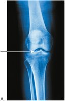
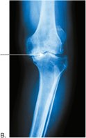
Symptoms include pain, stiffness, and reduced mobility.
Rheumatoid Arthritis
Rheumatoid arthritis is an autoimmune disorder causing chronic inflammation and deformity of joints.
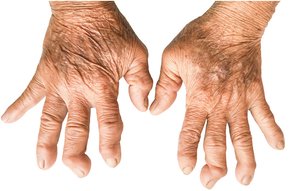
Commonly affects small joints of the hands and feet.
Laboratory, Diagnostic, and Radiologic Procedures
Bone Densitometry
Bone densitometry (DEXA scan) measures bone mineral density to assess osteoporosis risk.
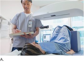
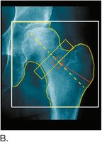
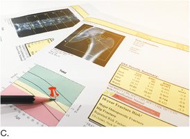
Common sites: hip, spine, wrist
Results help guide treatment decisions
Medical and Surgical Procedures
Fracture Management
Fractures are treated with immobilization (casts, splints), reduction (realignment), and sometimes surgery.
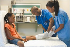
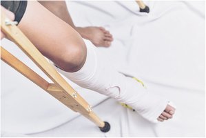
Physical therapy may be needed after healing to restore function.
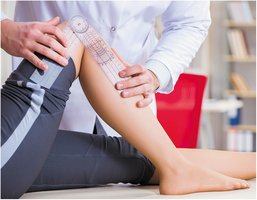
Range of Motion Assessment
Goniometers are used to measure joint range of motion, important in rehabilitation and diagnosis of joint disorders.
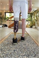
Prosthetics
Prosthetic limbs are custom-designed to replace lost extremities and restore mobility.
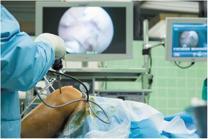
Arthroscopic Surgery
Arthroscopy is a minimally invasive surgical procedure for diagnosing and treating joint problems using a fiberoptic camera and instruments.
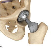
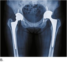
Hip Replacement
Total hip replacement involves removing the damaged femoral head and acetabulum and replacing them with prosthetic components.
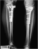
Internal Fixation
Severe fractures may require internal fixation with plates and screws to stabilize bone fragments during healing.
Summary Table: Major Bones of the Human Body
Region | Major Bones |
|---|---|
Skull | Frontal, parietal, temporal, occipital, sphenoid, ethmoid, maxilla, mandible, zygomatic |
Thorax | Sternum, ribs |
Vertebral Column | Cervical, thoracic, lumbar vertebrae, sacrum, coccyx |
Shoulder | Clavicle, scapula |
Arm | Humerus, radius, ulna |
Hand | Carpals, metacarpals, phalanges |
Pelvis | Ilium, ischium, pubis |
Leg | Femur, tibia, fibula, patella |
Foot | Tarsals, metatarsals, phalanges |
Key Medical Terminology
Oste/o-: bone
Arthr/o-: joint
Chondr/o-: cartilage
-itis: inflammation
-ectomy: surgical removal
-plasty: surgical repair
-scopy: visual examination
