 Back
BackIntroduction to Eukaryotic Cells: Structure, Function, and Classification
Study Guide - Smart Notes
 Tailored notes based on your materials, expanded with key definitions, examples, and context.
Overview of Eukaryotes
Endosymbiotic Theory and Evolution of Eukaryotes
The endosymbiotic theory explains the origin of eukaryotic cells as a result of sequential cell-merging events between ancient eukaryotic ancestors and certain prokaryotes. This theory is supported by evidence such as mitochondria and chloroplasts possessing their own circular DNA, 70S ribosomes, double-membrane structures, and the ability to replicate by binary fission.
Prokaryotes evolved ~3.5 billion years ago; eukaryotes evolved ~2.5 billion years ago.
Mitochondria originated from engulfed nonphotosynthetic prokaryotes.
Chloroplasts originated from engulfed photosynthetic prokaryotes (e.g., cyanobacteria).
Both organelles share similarities with bacteria, including gene structure and replication mechanisms.
Basic Description of Eukaryotic Cells and Differences from Prokaryotes
Eukaryotic cells are found in plants, animals, protists, and fungi. They are larger and more complex than prokaryotic cells, with larger genomes and multiple linear chromosomes. Eukaryotes possess a defined nucleus and a variety of membrane-bound organelles.
Eukaryotic cells: Larger, complex, multiple linear chromosomes, membrane-bound organelles.
Prokaryotic cells: Smaller, simpler, single circular chromosome, lack membrane-bound organelles.
Main Transport Mechanisms in Eukaryotic Cells
Eukaryotic cells utilize specialized transport mechanisms for importing and exporting substances:
Endocytosis: Imports substances into the cell by folding the plasma membrane around them, forming vesicles.
Exocytosis: Exports substances out of the cell via vesicles that fuse with the plasma membrane.
Types of endocytosis: Pinocytosis (cell drinking), Phagocytosis (cell eating), Receptor-mediated endocytosis.
Phagocytosis
Phagocytes (e.g., macrophages) engulf targets, forming a phagosome.
Phagosome fuses with lysosome, forming a phagolysosome where hydrolytic enzymes destroy most cells and viruses.
Waste products are expelled from the cell.
Classification of Eukaryotes
Four Kingdoms of Eukaryotes
Eukaryotic organisms are classified into four kingdoms: Animals, Plants, Fungi, and Protists. Each kingdom exhibits unique structural and functional characteristics.
Animals: Multicellular, do not carry out photosynthesis, obtain organic carbon from nutrients. Includes parasitic worms (helminths) and arthropods.
Plants: Multicellular, carry out photosynthesis, contain chloroplasts, serve as vehicles for infectious pathogens.
Fungi: Mostly multicellular, yeasts are unicellular, do not carry out photosynthesis, absorb nutrients, include pathogens and saprobes.
Protists: Diverse group, unicellular or multicellular, autotrophs or heterotrophs, asexual and sexual reproduction, may have cell walls.
Animals: Parasitic Helminths
Helminths are parasitic worms with complex life cycles, often spread in microscopic form. Examples include roundworms, flatworms, hookworms, tapeworms, and liver flukes.
WHO estimates half the world’s population is infected with some type of helminth.
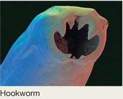
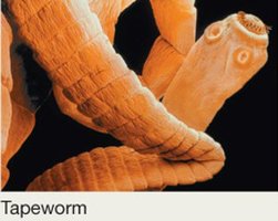
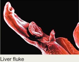
Fungi: Structure and Classification
Fungi grow as collections of tubular structures called hyphae. There are two types: septate hyphae (divided into individual cells) and aseptate hyphae (continuous chain with many nuclei). Many pathogenic fungi are dimorphic, cycling between hyphal and yeast-like forms.
Fungal spores help classify fungi and are either asexual (arise from mitosis) or sexual (arise from meiosis).
Asexual spores: conidiospores, sporangiospores.
Sexual spores: zygospores, ascospores, basidiospores.
Fungal Diseases (Mycoses)
Mycoses occur mainly in immunocompromised individuals or those with disrupted microbiota.
True pathogens infect healthy hosts (e.g., Histoplasmosis, Coccidioidomycosis).
Dermatophytes infect skin, hair, and nails (tinea infections).
Some fungi produce mycotoxins (e.g., ergot toxin from Claviceps purpurea).
Protists and Protozoans
Protists are a diverse group, including algae, slime molds, and protozoans. Protozoans are classified based on their motility:
Amoeboid: Use pseudopods (e.g., Entamoeba histolytica).
Flagellated: Use flagella (e.g., Trichomonas vaginalis, Giardia lamblia).
Ciliated: Use cilia (e.g., Balantidium coli).
Spore-forming (Apicomplexa): Move by gliding, obligate intracellular parasites, complex life cycles (e.g., Toxoplasma gondii, malaria).
Extracellular Structure
Plasma Membrane and Cell Wall
All eukaryotes have a plasma membrane composed of a phospholipid bilayer with sterols. Certain eukaryotes (fungi, plants, some protists) have a cell wall external to the plasma membrane, which lacks peptidoglycan and provides structural support.
Glycocalyx
Most eukaryotes possess a sticky extracellular layer called the glycocalyx, composed of carbohydrates, glycoproteins, and glycolipids. It functions in protection, adhesion, and cell recognition.
Flagella and Cilia
Eukaryotic flagella: Made of tubulin, 9+2 arrangement, anchored by basal body, wavelike motion.
Cilia: Structurally similar to flagella, shorter and more numerous, facilitate movement and environmental sensing.
Intracellular Structures
Ribosomes
Eukaryotic ribosomes are 80S, composed of 40S and 60S subunits.
Can be free in cytoplasm or bound to endoplasmic reticulum.
Mitochondria and chloroplasts contain 70S ribosomes, similar to prokaryotes.
Cytoskeleton
Dynamic network of protein fibers: microtubules, intermediate filaments, microfilaments.
Functions: Maintains shape, facilitates movement, protects, directs transport, coordinates cell division.
Microtubules
Hollow tubes (25 nm), made of tubulin, form spindle during cell division, arise from centrosome (two centrioles).
Intermediate Filaments
Rope-like fibers (10 nm), provide tensile strength.
Microfilaments
Fine fibers (3–6 nm), made of actin, associate with myosin, involved in muscle contraction and cell movement.
Membrane-Bound Organelles
Nucleus: Houses DNA, nucleolus for ribosome assembly, enclosed by nuclear envelope with pores.
Endoplasmic Reticulum (ER): Rough ER (protein modification), smooth ER (lipid production, detoxification).
Golgi Apparatus: Modifies, builds, sorts, and distributes proteins and lipids.
Vesicles: Transport, secretion, digestion (lysosomes, peroxisomes).
Vacuoles: Large sacs, mainly water and substances, regulate osmotic pressure in some protists.
Mitochondria and Chloroplasts
Double-membrane structures, 70S ribosomes, circular chromosome.
Mitochondria: ATP production, amino acid/vitamin synthesis, cell division regulation, apoptosis.
Structure: Smooth outer membrane, inner membrane with cristae, matrix.
Number varies by cell activity; most eukaryotic cells have mitochondria.
Chloroplasts: Photosynthetic cells only; structure includes inner/outer membrane, thylakoids (grana), stroma.
Summary Table: Eukaryotic vs. Prokaryotic Cells
Feature | Eukaryotic Cells | Prokaryotic Cells |
|---|---|---|
Nucleus | Present | Absent |
Membrane-bound organelles | Present | Absent |
Chromosomes | Multiple, linear | Single, circular |
Cell size | Larger | Smaller |
Ribosomes | 80S (cytoplasm), 70S (mitochondria/chloroplasts) | 70S |
Cell wall | Present in some (no peptidoglycan) | Present (peptidoglycan) |
Summary Table: Main Classes of Fungal Spores
Type | Origin | Examples |
|---|---|---|
Asexual | Mitosis | Conidiospores, Sporangiospores |
Sexual | Meiosis | Zygospores, Ascospores, Basidiospores |
Summary Table: Protozoan Groupings
Grouping | Motility Mechanism | Examples |
|---|---|---|
Amoeboid | Pseudopods | Entamoeba histolytica |
Flagellated | Flagella | Trichomonas vaginalis, Giardia lamblia |
Ciliated | Cilia | Balantidium coli |
Spore-forming | Gliding | Toxoplasma gondii, Plasmodium spp. |
Key Equations
Binary Fission (Prokaryotic and Organelle Replication):
ATP Production (Mitochondria):
Photosynthesis (Chloroplasts):
Additional info: Tables and equations were expanded for academic completeness and clarity. Images included are directly relevant to the explanation of helminth structure and classification.
