 Back
BackViruses, Viroids, and Prions: Structure, Classification, and Replication
Study Guide - Smart Notes
 Tailored notes based on your materials, expanded with key definitions, examples, and context.
Viruses, Viroids, and Prions
General Characteristics of Viruses
Viruses are unique infectious agents that differ fundamentally from bacteria and other microorganisms. They are obligate intracellular parasites, meaning they require living host cells to multiply. Viruses contain either DNA or RNA as their genetic material, but never both, and are surrounded by a protein coat called a capsid. They lack ribosomes and ATP-generating mechanisms, relying entirely on the host cell's machinery for replication.
Obligate intracellular parasites: Cannot reproduce outside a living cell.
Genetic material: DNA or RNA, single- or double-stranded, linear, circular, or segmented.
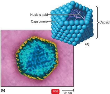
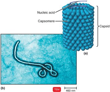
Capsid: Protein coat made of capsomeres.
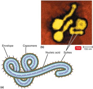
Envelope: Some viruses have an outer lipid, protein, and carbohydrate envelope derived from the host cell membrane.
No ribosomes or ATP-generating mechanisms.
Feature | Bacteria | Viruses |
|---|---|---|
Intracellular Parasite | No | Yes |
Plasma Membrane | Yes | No |
Binary Fission | Yes | No |
DNA and RNA | Yes | No (one or the other) |
ATP-Generating Metabolism | Yes | No |
Ribosomes | Yes | No |
Sensitivity to Antibiotics | Yes | No |
Sensitivity to Interferon | No | Yes |
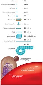
Host Range: The spectrum of host cells a virus can infect is determined by specific attachment sites and cellular factors. Most viruses infect only specific types of cells in one host species.
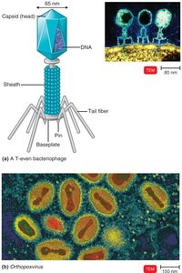
Viral Structure
The complete, fully developed infectious viral particle is called a virion. Viruses exhibit diverse structures, which can be classified based on their morphology:
Helical viruses: Hollow, cylindrical capsid (e.g., rabies, Ebola).
Polyhedral viruses: Many-sided, usually icosahedral (e.g., adenoviruses, poliovirus).
Enveloped viruses: Spherical, with a lipid envelope (e.g., influenza, herpesviruses).
Complex viruses: Complicated structures, such as bacteriophages.
Taxonomy and Classification of Viruses
Viruses are classified based on their nucleic acid type, replication strategy, and morphology. The Baltimore classification system groups viruses according to their genome type and method of mRNA production. Viral taxonomy uses the following conventions:
Order names: End in -virales
Family names: End in -viridae
Genus names: End in -virus
Viral species: Group of viruses sharing the same genetic information and ecological niche (host)
Isolation, Cultivation, and Identification of Viruses
Viruses must be grown in living cells. Methods include:
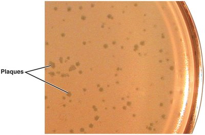
Bacteriophages: Grown in bacteria; form plaques on bacterial lawns, each representing a single virus.
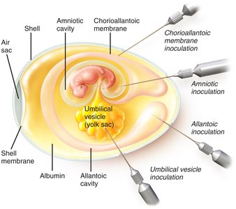
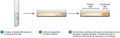
Animal viruses: Grown in living animals, embryonated eggs, or cell cultures (primary, diploid, or continuous cell lines).
Viral identification methods include observation of cytopathic effects, serological tests (e.g., ELISA), and nucleic acid-based tests (e.g., PCR).
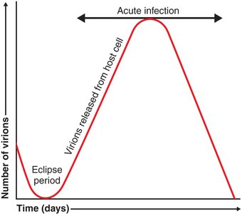
Viral Multiplication
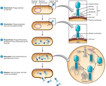
Viruses multiply by taking over the host cell's metabolic machinery. The process differs between bacteriophages and animal viruses.
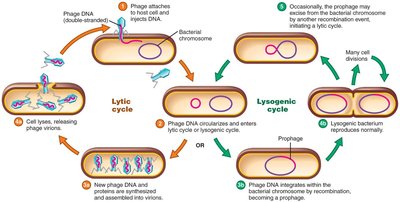
Lytic cycle (bacteriophages): Virus causes lysis and death of the host cell.
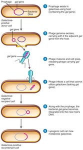
Lysogenic cycle (bacteriophages): Viral DNA integrates into the host genome as a prophage and can remain latent.
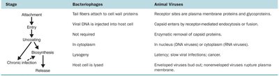
Stage | Bacteriophages | Animal Viruses |
|---|---|---|
Attachment | Tail fibers attach to cell wall proteins | Attachment sites are plasma membrane proteins and glycoproteins |
Entry | Viral DNA is injected into host cell | Capsid enters by receptor-mediated endocytosis or fusion |
Uncoating | Not required | Enzymatic removal of capsid proteins |
Biosynthesis | In cytoplasm | In nucleus (DNA viruses) or cytoplasm (RNA viruses) |
Chronic Infection | Lysogeny | Latency; slow viral infections; cancer |
Release | Host cell is lysed | Enveloped viruses bud out; nonenveloped viruses rupture plasma membrane |
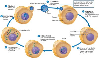
Multiplication of Animal Viruses
The general steps for animal virus replication are:
Attachment: Virus attaches to host cell membrane.
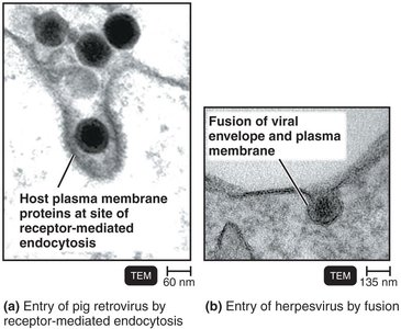
Entry: By receptor-mediated endocytosis or fusion.
Uncoating: Separation of viral nucleic acid from capsid by viral or host enzymes.
Biosynthesis: Production of viral nucleic acid and proteins.
Maturation: Assembly of viral components.
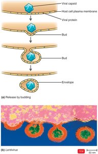
Release: By budding (enveloped viruses) or rupture (nonenveloped viruses).
Biosynthesis of DNA and RNA Viruses
DNA viruses generally replicate their DNA in the nucleus and synthesize capsid proteins in the cytoplasm. RNA viruses replicate in the cytoplasm using RNA-dependent RNA polymerase. Retroviruses use reverse transcriptase to produce DNA from their RNA genome.
Viruses and Cancer
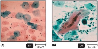
Some viruses can cause cancer by integrating their genetic material into the host genome, leading to transformation of normal cells into tumor cells. Oncogenes are mutated proto-oncogenes that drive uncontrolled cell growth. Oncogenic viruses include members of the Herpesviridae, Papovaviridae, Hepadnaviridae, and Retroviridae families.
DNA oncogenic viruses: HPV (cervical cancer), Epstein-Barr virus (Burkitt's lymphoma), Hepatitis B virus (liver cancer).
RNA oncogenic viruses: Retroviruses (HTLV-1, HTLV-2, HIV).
Latent and Persistent Viral Infections
Latent infections are characterized by periods of inactivity, with the virus remaining in the host cell without producing disease (e.g., herpes simplex virus, varicella-zoster virus). Persistent infections involve continuous viral replication and release, often leading to chronic disease (e.g., HIV, hepatitis B).
Plant Viruses and Viroids
Plant viruses typically enter host cells through wounds or via insect vectors. Viroids are infectious RNA molecules lacking a protein coat, causing diseases such as potato spindle tuber disease. Virusoids are viroids enclosed in a protein coat, requiring coinfection with a virus to cause disease.
Prions
Prions are infectious proteins that cause neurodegenerative diseases by inducing abnormal folding of normal cellular proteins. Prion diseases include Creutzfeldt-Jakob disease, mad cow disease, and scrapie. The infectious form, PrPSc, converts normal PrPC into the abnormal form, leading to accumulation and cell death.
Summary Table: Key Differences Between Viruses, Viroids, and Prions
Agent | Genetic Material | Protein Coat | Example Disease |
|---|---|---|---|
Virus | DNA or RNA | Present | Influenza, HIV, Herpes |
Viroid | RNA (short, circular) | Absent | Potato spindle tuber disease |
Prion | None (protein only) | Absent | Creutzfeldt-Jakob disease |
