Back
BackEukaryotic Cell Structure and Function: Biochemistry Study Guide
Study Guide - Smart Notes
 Tailored notes based on your materials, expanded with key definitions, examples, and context.
Eukaryotic Cell Structure and Function
Common Features of Eukaryotic Cells
Eukaryotic cells are distinguished by their structural complexity and larger size compared to bacterial and archaeal cells. They possess membrane-bound organelles that compartmentalize cellular functions, allowing for greater specialization and efficiency.
Membrane-delimited nucleus: Houses genetic material and separates it from the cytoplasm.
Membrane-bound organelles: Includes mitochondria, endoplasmic reticulum, Golgi apparatus, lysosomes, and others.
Intracytoplasmic membrane complex: Facilitates transport within the cell.
Defined nucleus: The presence of a nucleus is a hallmark of eukaryotes.
Eukaryotic Organelles and Their Functions
Eukaryotic cells contain specialized organelles, each with distinct roles in cellular metabolism, genetic regulation, and energy production.
Nucleus: Contains DNA attached to histone proteins; site of genetic regulation.
Nucleolus: Not membrane-bound; directs synthesis and processing of rRNA for ribosome assembly.
Ribosomes: 80S in size (60S + 40S subunits); may be free or attached to ER; responsible for protein synthesis.
Mitochondria: Site of TCA cycle, electron transport, and ATP generation via oxidative phosphorylation.
Hydrogenosomes: Energy conservation organelles in some anaerobic protists; generate ATP by fermentation.
Chloroplasts: Site of photosynthesis; contains thylakoids and stroma for light and dark reactions.
Endoplasmic Reticulum (ER): Rough ER synthesizes proteins; Smooth ER synthesizes lipids.
Golgi Apparatus: Modifies, packages, and secretes materials.
Lysosomes: Contain digestive enzymes for hydrolysis of macromolecules.
Peroxisomes: Oxidize compounds and degrade hydrogen peroxide.
Cilia and Flagella: Provide motility; contain microtubules in a 9+2 arrangement.
Cell Membranes: Lipid bilayer with phosphoglycerides, sphingolipids, and cholesterol.
Cell Walls: Composition varies; fungi (chitin), plants (cellulose), protozoa (absent or glycoprotein).
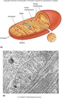
Mitochondria
Mitochondria are the "powerhouses" of the cell, responsible for energy production through the TCA cycle and oxidative phosphorylation. They have a double membrane structure, with the inner membrane forming cristae to increase surface area for metabolic reactions.
Outer membrane: Contains porin proteins.
Inner membrane: Highly folded (cristae); site of electron transport chain.
Matrix: Contains mitochondrial DNA, ribosomes, and enzymes for the TCA cycle.
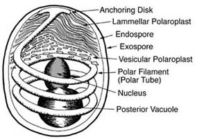
Microsporidia
Microsporidia are obligate intracellular parasites notable for their lack of mitochondria. They possess unique structures such as polar filaments and vacuoles, and may represent advanced prokaryotes.
No mitochondria: Energy metabolism differs from typical eukaryotes.
Polar filament: Used for infection of host cells.
Complex spore structure: Includes anchoring disk, polaroplasts, and vacuoles.
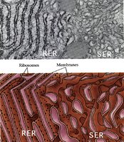
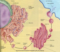
Endoplasmic Reticulum (ER)
The ER is a network of membranous tubules and sacs involved in synthesis and transport of proteins and lipids. It is divided into rough and smooth regions based on the presence of ribosomes.
Rough ER (RER): Ribosomes attached; synthesizes secreted proteins.
Smooth ER (SER): Lacks ribosomes; synthesizes lipids.
Functions: Transports proteins, lipids, and other materials; major site of membrane synthesis.
Golgi Apparatus
The Golgi apparatus is a stack of cisternae involved in the modification, packaging, and secretion of cellular products. It has distinct cis and trans faces for receiving and dispatching vesicles.
Dictyosomes: Stacks of cisternae.
Modification: Glycosylation and other post-translational changes.
Packaging: Formation of secretory vesicles.
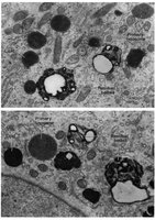
Lysosomes
Lysosomes are membrane-enclosed organelles containing hydrolytic enzymes for digestion of macromolecules. They maintain an acidic internal pH and isolate lytic activity from the rest of the cell.
Digestive enzymes: Hydrolyze proteins, lipids, nucleic acids.
Residual bodies: Contain indigestible material.
Internal pH: Approximately 5.
Endocytic Pathways
Endocytosis is the process by which eukaryotic cells internalize materials from their environment. It involves the formation of vesicles from the plasma membrane and delivery of contents to lysosomes for degradation.
Phagocytosis: Engulfment of large particles.
Clathrin-dependent: Protein-coated pits bind specific macromolecules.
Caveolin-dependent: Involved in signal transduction and transport.
Endosomes: Intermediate organelles in the pathway to lysosomes.
Peroxisomes
Peroxisomes are small organelles that oxidize various compounds, producing hydrogen peroxide, which is then degraded by catalase. They play a role in detoxification and lipid metabolism.
Oxidation reactions: Alcohols, fatty acids, toxins.
Catalase: Converts hydrogen peroxide to water and oxygen.
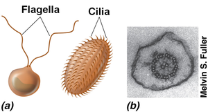
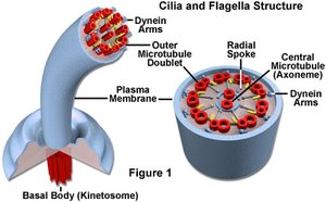
Cilia and Flagella
Cilia and flagella are motile structures composed of microtubules arranged in a 9+2 pattern. They are involved in cell movement and fluid transport across cell surfaces.
Flagella: Long, whip-like; undulating movement.
Cilia: Short, oar-like; coordinated beating.
Axoneme: Core structure with microtubules.
Basal body: Directs synthesis and anchoring.
Eukaryotic Cell Membranes
The plasma membrane of eukaryotic cells is a lipid bilayer composed of phosphoglycerides, sphingolipids, and cholesterol. These components contribute to membrane strength and fluidity, and microdomains facilitate various cellular processes.
Lipid composition: More similar to bacteria than archaea.
Microdomains: Specialized regions for signaling and transport.
Eukaryotic Cell Walls
Cell walls in eukaryotes vary in composition and presence. Fungi have chitin or glucans, plants have cellulose, protozoa may lack cell walls, and animals have an extracellular matrix instead.
Fungi: Chitin or glucans.
Algae/Plants: Cellulose (diatoms: silica).
Protozoa: Absent or glycoprotein in spore/cyst stage.
Animals: No cell wall; extracellular matrix present.
Eukaryotic Cytoskeleton and Cytoplasm
The cytoskeleton provides structural support and facilitates intracellular transport. It consists of actin filaments, intermediate filaments, and microtubules.
Actin filaments: Support cell shape and movement.
Intermediate filaments: Provide mechanical strength.
Microtubules: Involved in cell division and transport.
Comparison of Bacterial, Archaeal, and Eukaryotic Cells
Eukaryotic cells differ from bacterial and archaeal cells in their complexity, size, and presence of a nucleus. However, all three domains share fundamental biochemical processes and the genetic code.
Nucleus: Present only in eukaryotes.
Cell size: Eukaryotes are generally larger.
Cell division: Eukaryotes undergo mitosis and meiosis.
Molecular unity: Biochemical pathways and genetic code are conserved.
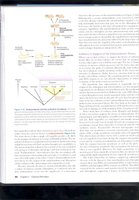
Endosymbiotic Theory and Origin of Eukaryotic Cells
The endosymbiotic theory explains the origin of mitochondria and chloroplasts as a result of symbiotic relationships between ancestral eukaryotic cells and bacteria. Evidence includes similarities in structure, DNA, and division mechanisms.
Double membrane: Consistent with engulfment theory.
Circular DNA: Similar to bacterial genomes.
Independent division: Binary fission, like bacteria.
Ribosomes: 70S, similar to bacteria.
Modern Endosymbiosis
Contemporary examples of endosymbiosis can be observed in amoeba and x-bacteria, providing experimental evidence for the theory.